1206711-16-1
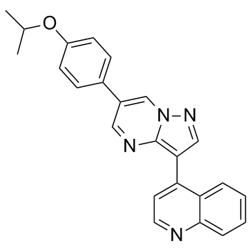
1206711-16-1 structure
- Name: DMH-1
- Chemical Name: 4-[6-(4-propan-2-yloxyphenyl)pyrazolo[1,5-a]pyrimidin-3-yl]quinoline
- CAS Number: 1206711-16-1
- Molecular Formula: C24H20N4O
- Molecular Weight: 380.442
- Catalog: Biochemical Inhibitor TGF-beta/Smad signaling pathway inhibitor (TGF-beta/Smad) TGF-beta/Smad inhibitor
- Create Date: 2016-08-19 22:27:47
- Modify Date: 2026-07-01 11:34:22
-
DMH-1 is a potent and selective BMP inhibitor with IC50s of 27/107.9/<5/47.6 nM for ALK1/ALK2/ALK3/ALK6, respectively.
| Name | 4-[6-(4-propan-2-yloxyphenyl)pyrazolo[1,5-a]pyrimidin-3-yl]quinoline |
|---|---|
| Synonyms |
4-[6-(4-Isopropoxyphenyl)pyrazolo[1,5-a]pyrimidin-3-yl]quinoline
DMH1 DMH-1 |
| Description | DMH-1 is a potent and selective BMP inhibitor with IC50s of 27/107.9/<5/47.6 nM for ALK1/ALK2/ALK3/ALK6, respectively. |
|---|---|
| Related Catalog | |
| Target |
IC50: 27 nM (ALK1), 107.9 nM (ALK2), <5 nM (ALK3), 47.6 nM (ALK6)[1] |
| In Vitro | DMH-1 (0.5 μM) induces regulation of OCT4, Nanog, and PAX6 protein expression. DMH-1 significantly reduces the percentage of cells expressing the pluripotency marker proteins OCT4 and Nanog in both SM3 and CA6 cells. PAX6 expression is significantly up-regulated by day 5 and day 7 in CA6 and SM3 cells, respectively. DMH-1 induces regulation of pluripotency and neural precursor marker mRNAs. PAX6 can regulate the expression of SOX1 independently by manipulating the DMH-1 concentration during the neural induction of hiPSCs[2]. DMH-1 (5 μM and 10 μM) inhibits CDDP-induced autophagy in HeLa cells and enhances the ability of CDDP to reduce HeLa cell viability, inhibits tamoxifen-induced autophagy in MCF-7 cells and enhances the ability of tamoxifen to reduce MCF-7 cell viability, inhibits 5-FU-induced autophagy in both MCF-7 and HeLa cells but does not affect the inhibitory effects of 5-FU on MCF-7 and HeLa cell viability. DMH-1 enhances the apoptotic induction effects of CDDP on HeLa cells after 24 h treatment. DMH-1 inhibits HeLa and MCF-7 cell proliferation[3]. DMH-1 (20 μM) reduces the canonical phosphorylation of Smads 1,5 and 9. DMH-1 in combination with Cisplatin significantly decreases Ki-67 positive staining in the OVCAR8 cells. DMH-1 (20 µM) upregulates JAG1, reduces CYP1B1 and increases HAPLN1 expression in both OVCAR8 and NCI-RES cells[4]. |
| In Vivo | DMH1 (5 mg/kg, i.p.) treatment significantly reduces the tumor growth in human lung cancer xenograft model[5]. |
| Cell Assay | Cells are seeded in 96-well plates and treated with different drugs for appropriate time. Then 5 mg/mL MTT is added and incubated for 4 h at 37°C. Medium is then removed and 200 μL of DMSO is added to dissolve the crystal. Absorbance is measured at a wavelength of 490 nm with a plate reader. |
| Animal Admin | Sub-confluent A549 cells are trypsinized and then suspended in serum free RPMI 1640 medium. The cell suspension (1×10⁶ cells in 100 µL medium for each injection) is injected subcutaneously into both the right and left flanks of eight-week old NOD SCID mice (n=5 for each group). Mice are given Intraperitoneal (i.p.) injection of the vehicle (12.5% 2-hydroxypropyl-β-cyclodextrin) or 5 mg/kg DMH1 every other day. The tumor sizes are measured with a vernier caliper from the sixth day to the fourth week after tumor implantation. The tumor volume (V) is calculated according to the formulation: Volume=(width)2×length/2. The tumor tissues are dissected at the end of study, and are sectioned and stained with H & E, and for immunohistochemical analysis. |
| References |
| Density | 1.2±0.1 g/cm3 |
|---|---|
| Molecular Formula | C24H20N4O |
| Molecular Weight | 380.442 |
| Exact Mass | 380.163696 |
| PSA | 52.31000 |
| LogP | 3.62 |
| Appearance | light yellow to orange |
| Index of Refraction | 1.672 |
| Storage condition | Store at +4°C |
| Water Solubility | DMSO: soluble5mg/mL, clear (warmed) |
| Symbol |

GHS06 |
|---|---|
| Signal Word | Danger |
| Hazard Statements | H301 |
| Precautionary Statements | P301 + P310 |
| Hazard Codes | T |
| Risk Phrases | 25 |
| Safety Phrases | 45 |
| RIDADR | UN 2811 6.1 / PGIII |