Janus Green B
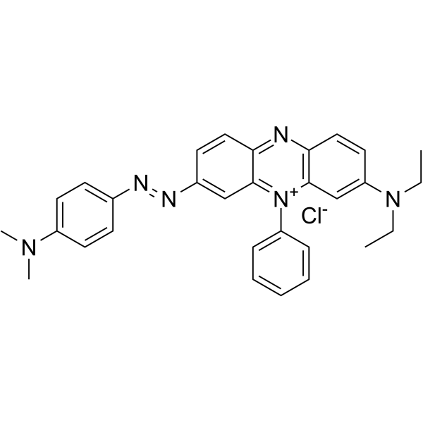
Janus Green B structure
|
Common Name | Janus Green B | ||
|---|---|---|---|---|
| CAS Number | 2869-83-2 | Molecular Weight | 511.060 | |
| Density | N/A | Boiling Point | N/A | |
| Molecular Formula | C30H31ClN6 | Melting Point | 160 °C (dec.)(lit.) | |
| MSDS | USA | Flash Point | N/A | |
|
Induction of apoptotic cell death by betulin in multidrug-resistant human renal carcinoma cells.
Oncol. Rep. 34 , 1058-64, (2015) Betulin, a triterpene from the bark of various species of birch tree, has various biological effects, including antiviral, antifungal and anticancer activities. The aim of the present study was to elucidate the mechanisms underlying the apoptotic effect of be... |
|
|
Analysis and testing of biological stains--the Biological Stain Commission Procedures.
Biotech. Histochem. 77(5&6) , 237-275, (2002)
|
|
|
Complete staining of human spermatozoa and immature germ cells combined with phase contrast microscopy.
Arch. Androl. 19(3) , 217-21, (1987) A method combining Janus green B and Thymol blue stains the anterior part of the head, the nuclear membrane, middle piece, and tail of spermatozoa light green and the nucleus deep purple. The method provides excellent stained preparations for the evaluation o... |
|
|
Determination of viability of Histoplasma capsulatum yeast cells grown in vitro: comparison between dye and colony count methods.
J. Med. Vet. Mycol. 25(2) , 107-14, (1987) The viability of Histoplasma capsulatum yeast cells grown under different conditions was determined by dye tests with Eosin-Y and Janus Green B and by colony counts of cells plated on brain-heart infusion agar supplemented with histoplasma growth factor and b... |
|
|
Flow cytometric identification of two different rhodamine-123-stained mitochondrial populations in maize leaves.
Protoplasma 231(3-4) , 249-52, (2007) Flow cytometric analysis of mitochondria isolated from maize leaves revealed two distinct rhodamine-123-stained fluorescence populations distinguishable by their main fluorescence channel. Further microscopic observation of mitochondria stained with Janus Gre... |
|
|
Long wavelength fluorescence lifetime standards for front-face fluorometry.
J. Fluoresc. 20(2) , 435-40, (2010) With the increased development and use of fluorescence lifetime-based sensors, fiber optic sensors, fluorescence lifetime imaging microscopy (FLIM), and plate and array readers, , calibration standards are essential to ensure the proper function of these devi... |
|
|
Regression of the interdigital tissue during the formation of the digits.
Acta Histochem. Suppl. 32 , 165-9, (1986)
|
|
|
René Couteaux (1909-1999) and the morphological identification of synapses.
Biol. Cell 98(8) , 503-9, (2006) During a historical research, we realized that René Couteaux (1909-1999) was the first histologist who stained the postsynaptic structure of the neuromuscular junction. By means of Janus Green B dye, he revealed the membranous 'subneural apparatus' related to... |
|
|
Reduction of organic dyes in matrix-assisted laser desorption/ionization and desorption/ionization on porous silicon.
Rapid Commun. Mass Spectrom. 18(23) , 2811-7, (2004) Reduction of analytes in matrix-assisted laser desorption/ionization (MALDI) often obscures the actual determination of molecular structure. To address the redox reactions in laser desorption/ionization processes, the organic dyes Methylene Blue, Janus Green ... |
|
|
Janus Green B as a rapid, vital stain for peripheral nerves and chordotonal organs in insects.
J. Neurosci. Methods 49(1-2) , 17-22, (1993) Effective staining of peripheral nerves in live insects is achieved with the vital stain Janus Green B. A working solution of 0.02% Janus Green B in saline is briefly applied to the exposed peripheral nervous system. The stain is then decanted and the dissect... |