5-Iodouracil
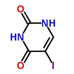
5-Iodouracil structure
|
Common Name | 5-Iodouracil | ||
|---|---|---|---|---|
| CAS Number | 696-07-1 | Molecular Weight | 237.983 | |
| Density | 2.4±0.1 g/cm3 | Boiling Point | 401ºC | |
| Molecular Formula | C4H3IN2O2 | Melting Point | 274-276 °C (dec.)(lit.) | |
| MSDS | Chinese USA | Flash Point | N/A | |
| Symbol |

GHS07 |
Signal Word | Warning | |
|
Enzymatic primer-extension with glycerol-nucleoside triphosphates on DNA templates.
PLoS ONE 4(3) , e4949, (2009) Glycerol nucleic acid (GNA) has an acyclic phosphoglycerol backbone repeat-unit, but forms stable duplexes based on Watson-Crick base-pairing. Because of its structural simplicity, GNA is of particular interest with respect to the possibility of evolving func... |
|
|
Highly hydrophilic and nonionic poly(2-vinyloxazoline)-grafted silica: a novel organic phase for high-selectivity hydrophilic interaction chromatography.
Anal. Bioanal. Chem 406(19) , 4585-93, (2014) A new hydrophilic and nonionic poly(2-vinyloxazoline)-grafted silica (Sil-VOX(n)) phase was synthesized and applied for the separation of nucleosides and nucleobases in hydrophilic interaction chromatography (HILIC). Polymerization and immobilization onto sil... |
|
|
Preparation and properties of oligodeoxynucleotides containing 5-iodouracil and 5-bromo- and 5-iodocytosine.
Bioconjug. Chem. 8(5) , 757-61, (1997) The behavior of oligonucleotides containing 5-iodouracil, 5-bromocytidine, and 5-iodocytidine in concentrated ammonia is described. 5-Aminouracil and 5-aminocytidine are obtained as side products when deprotection is performed at 60 degrees C. Small amounts, ... |
|
|
A simple crosslinking method, CLAMP, to map the sites of RNA-contacting domains within a protein.
Methods Mol. Biol. 488 , 181-90, (2008) A large number of proteins contain multiple RNA recognition motifs (RRMs). How multiple RRMs contribute to RNA recognition in solution is, however, poorly understood. Here, we describe a simple biochemical approach called CLAMP (crosslinking and mapping of pr... |
|
|
Photoreactivity of 5-iodouracil-containing DNA-Sso7d complex.
Nucleic Acids Symp. Ser. (42) , 171-2, (1999) X-ray structure of DNA-Sso7d complex indicated that binding of this protein causes sharp DNA bending. In order to examine whether this protein also causes DNA bending in solution, photoreactions of 1U-substituted DNA in the presence and the absence of Sso7d p... |
|
|
Photocrosslinking of 5-iodouracil-substituted RNA and DNA to proteins.
Science 262(5137) , 1255-7, (1993) 5-Iodouracil-substituted RNA and DNA were crosslinked regiospecifically to associated proteins in yields of 70 to 94% of bound nucleic acid. Irradiation of the iodouracil chromophore with monochromatic, long-wavelength ultraviolet radiation (325 nanometers) e... |
|
|
Efficient C2'alpha-hydroxylation of deoxyribose in protein-induced Z-form DNA.
J. Am. Chem. Soc. 125(6) , 1526-31, (2003) DNA local conformations are thought to play an important biological role in processes such as gene expression by altering DNA-protein interactions. Although left-handed Z-form DNA is one of the best-characterized and significant local structures of DNA, havin... |
|
|
Photoreactivity of 5-iodouracil-containing DNA-Sso7d complex in solution: the protein-induced DNA kink causes intrastrand hydrogen abstraction from the 5-methyl of thymine at the 5' side.
J. Am. Chem. Soc. 124(10) , 2086-7, (2002) Photoirradiation of 5-iodouracil-containing DNA, d(GTAAT(I)UAC)(2) with Sso7d protein, possessing significant kink in DNA in the crystal structure induces an unprecedented intrastrand H abstraction at the methyl group of T(5), together with selective photooxi... |
|
|
Electrospray ionization mass spectrometric characterization of photocrosslinked DNA-EcoRI DNA methyltransferase complexes.
Nucleic Acids Res. 26(2) , 645-9, (1998) We describe a novel strategy combining photocrosslinking and HPLC-based electrospray ionization mass spectrometry to identify UV crosslinked DNA-protein complexes. Eco RI DNA methyltransferase modifies the second adenine within the recognition sequence GAATTC... |
|
|
Mutational analysis of target base flipping by the EcoRV adenine-N6 DNA methyltransferase.
J. Mol. Biol. 285(3) , 1121-30, (1999) DNA methyltransferases flip their target base out of the DNA helix. Here, we have investigated base flipping by wild-type EcoRV DNA methyltransferase (M.EcoRV) and five M.EcoRV variants (D193A, Y196A, S229A, W231R and Y258A). These variants bind to DNA and S-... |