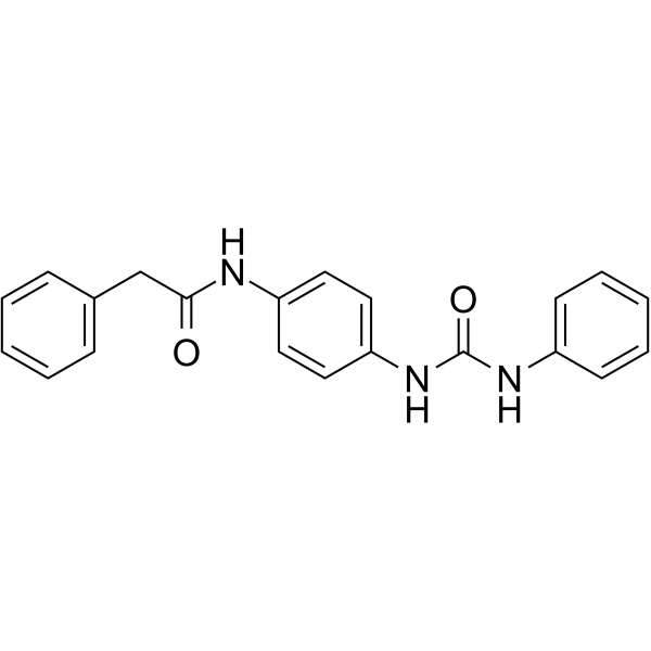
| In Vitro |
VEGFR-2-IN-19 (Compound 15b) (0-100 µM, 48 h) shows antiproliferative activity against cancer cells[1]. VEGFR-2-IN-19 (0-10 µM, 48 h) inhibits the phosphorylation of VEGFR2 and the Raf/MEK/ERK signaling pathway, induces cell apoptosis, and arrests cell cycle at G0/G1 phase in HT-29 cells[1]. VEGFR-2-IN-19 (0-10 µM, 12 h) increases intracellular reactive oxygen species level in HT-29 cells[1]. Cell Cytotoxicity Assay[1] Cell Line: HT-29 and A549 Concentration: 0-100 µM Incubation Time: 48 h Result: Showed antiproliferative activity with IC50 values of 3.38±1.3 µM and 0.63±0.04 µM against HT-29 and A549 cells, respectively. Western Blot Analysis[1] Cell Line: HT-29 Concentration: 0, 1, 5, and 10 µM Incubation Time: 48 h Result: Effectively inhibited the phosphorylation of VEGFR2 in dose-dependent manner. Suppressed dose-dependently expression of c-Raf, ERK1/2, and MEK and phosphorylation of ERK1/2 (pERK1/2). Up-regulated caspase-3 and Bax expression, down-regulated Bcl-2 expression. Apoptosis Analysis[1] Cell Line: HT-29 Concentration: 0, 1, 5, and 10 µM Incubation Time: 48 h Result: The total numbers of early and late apoptotic cells were increased significantly relative to that of untreated control cells in dose-dependent manner. Cell Cycle Analysis[1] Cell Line: HT-29 Concentration: 0, 1, 5, and 10 µM Incubation Time: 48 h Result: Increased the population of cells in sub G1 and G0/G1 phase and decreased the population of cells in S phase.
|
