A standardized method for the construction of tracer specific PET and SPECT rat brain templates: validation and implementation of a toolbox.
David Vállez Garcia, Cindy Casteels, Adam J Schwarz, Rudi A J O Dierckx, Michel Koole, Janine Doorduin
Index: PLoS ONE 10(3) , e0122363, (2015)
Full Text: HTML
Abstract
High-resolution anatomical image data in preclinical brain PET and SPECT studies is often not available, and inter-modality spatial normalization to an MRI brain template is frequently performed. However, this procedure can be challenging for tracers where substantial anatomical structures present limited tracer uptake. Therefore, we constructed and validated strain- and tracer-specific rat brain templates in Paxinos space to allow intra-modal registration. PET [18F]FDG, [11C]flumazenil, [11C]MeDAS, [11C]PK11195 and [11C]raclopride, and SPECT [99mTc]HMPAO brain scans were acquired from healthy male rats. Tracer-specific templates were constructed by averaging the scans, and by spatial normalization to a widely used MRI-based template. The added value of tracer-specific templates was evaluated by quantification of the residual error between original and realigned voxels after random misalignments of the data set. Additionally, the impact of strain differences, disease uptake patterns (focal and diffuse lesion), and the effect of image and template size on the registration errors were explored. Mean registration errors were 0.70 ± 0.32 mm for [18F]FDG (n = 25), 0.23 ± 0.10mm for [11C]flumazenil (n = 13), 0.88 ± 0.20 mm for [11C]MeDAS (n = 15), 0.64 ± 0.28 mm for [11C]PK11195 (n = 19), 0.34 ± 0.15 mm for [11C]raclopride (n = 6), and 0.40 ± 0.13 mm for [99mTc]HMPAO (n = 15). These values were smallest with tracer-specific templates, when compared to the use of [18F]FDG as reference template (p<0.001). Additionally, registration errors were smallest with strain-specific templates (p<0.05), and when images and templates had the same size (p ≤ 0.001). Moreover, highest registration errors were found for the focal lesion group (p<0.005) and the diffuse lesion group (p = n.s.). In the voxel-based analysis, the reported coordinates of the focal lesion model are consistent with the stereotaxic injection procedure. The use of PET/SPECT strain- and tracer-specific templates allows accurate registration of functional rat brain data, independent of disease specific uptake patterns and with registration error below spatial resolution of the cameras. The templates and the SAMIT package will be freely available for the research community [corrected].
Related Compounds
| Structure | Name/CAS No. | Molecular Formula | Articles |
|---|---|---|---|
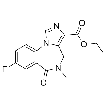 |
Flumazenil
CAS:78755-81-4 |
C15H14FN3O3 | |
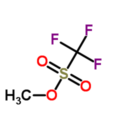 |
Methyl trifluoromethanesulfonate
CAS:333-27-7 |
C2H3F3O3S |
|
Tonic current through GABAA receptors and hyperpolarization-...
2014-07-01 [Eur. J. Neurosci. 40(1) , 2241-54, (2014)] |
|
Mild Staphylococcus aureus Skin Infection Improves the Cours...
2015-01-01 [PLoS ONE 10 , e0129150, (2015)] |
|
PET molecular imaging to investigate higher brain dysfunctio...
2013-01-01 [Acta Neurochir. Suppl. 118 , 251-4, (2013)] |
|
Cortical oscillatory dynamics and benzodiazepine-site modula...
2015-08-01 [Neuropharmacology 95 , 192-205, (2015)] |
|
Effect of small molecule vasopressin V1a and V2 receptor ant...
2015-02-15 [J. Neurotrauma 32(4) , 221-7, (2015)] |