| Structure | Name/CAS No. | Articles |
|---|---|---|
 |
Sodium hydroxide
CAS:1310-73-2 |
|
 |
Fluorescein sodium
CAS:518-47-8 |
|
 |
Glycine
CAS:56-40-6 |
|
 |
Disodium hydrogenorthophosphate
CAS:7558-79-4 |
|
 |
Citric Acid
CAS:77-92-9 |
|
 |
3-Ethyl-2,4-pentanedione
CAS:1540-34-7 |
|
 |
Acid Red 87
CAS:17372-87-1 |
|
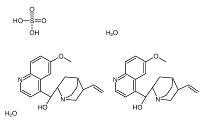 |
Quinine hemisulfate hydrate
CAS:207671-44-1 |
|
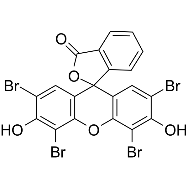 |
Eosin Y
CAS:15086-94-9 |
|
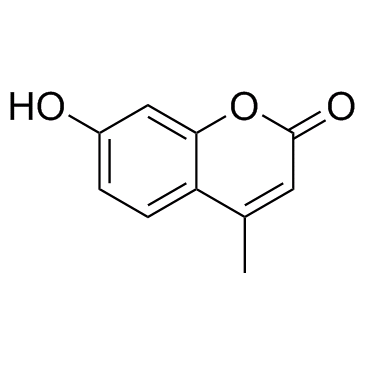 |
4-Methylumbelliferone
CAS:90-33-5 |