Materials Science and Engineering: C 2015-01-01

Water-soluble nanoconjugates of quantum dot-chitosan-antibody for in vitro detection of cancer cells based on "enzyme-free" fluoroimmunoassay.

Herman S Mansur, Alexandra A P Mansur, Amanda Soriano-Araújo, Zélia I P Lobato, Sandhra M de Carvalho, Maria de Fatima Leite

Index: Mater. Sci. Eng. C. Mater. Biol. Appl. 52 , 61-71, (2015)

Full Text: HTML

Abstract

Cancer remains one of the world's most devastating diseases with millions of fatalities and new cases every year. In this work, we attempted to develop a facile "enzyme-free" fluoroimmunoassay based on the novel nanoconjugates composed of CdS quantum dots (QDs) as the fluorescent inorganic core and an antibody-modified polysaccharide as the organic shell, modeling their possible application for the in vitro diagnosis of non-Hodgkin lymphoma (NHL) cancer. Chitosan was conjugated with an anti-CD20 polyclonal antibody (pAbCD20) by the formation of covalent amide bonds. In the sequence, these chitosan-antibody conjugates were utilized as direct ligands for the surface biofunctionalization of CdS QDs (CdS/chitosan-pAbCD20) using a single-step colloidal process in aqueous medium at room temperature. The most relevant physico-chemical properties of these nanoconjugates were assessed by morphological and spectroscopic techniques. The results indicated that CdS nanocrystals were produced with an average diameter of 2.5 nm and with cubic zinc blende crystalline nanostructure. The CdS-immunoconjugates (CdS/chitosan-pAbCD20) presented colloidal hydrodynamic diameter (HD) of 15.0 ± 1.2n m. In addition, the results evidenced that the "enzyme-free" QD-linked immunosorbent assay (QLISA) was effective for the in vitro detection against the antigen CD20 (aCD20) based on fluorescent behavior of the CdS nanoconjugates. Moreover, the CdS-immunoconjugates were successfully used for fluorescence bioimaging of NHL cancer cells. Finally, the cell viability results using different cell cultures based on LDH, MTT and Resazurin bio-assays have demonstrated no cytotoxicity of the new CdS-chitosan bioconjugates relative to the standard controls. Thus, CdS conjugates may offer a promising platform for the future development of in vitro and in vivo applications for the detection and diagnosis of NHL cancer cells.Copyright © 2015 Elsevier B.V. All rights reserved.

Related Compounds

Structure Name/CAS No. Articles
1-(3-Dimethylaminopropyl)-3-ethylcarbodiimide hydrochloride Structure 1-(3-Dimethylaminopropyl)-3-ethylcarbodiimide hydrochloride
CAS:25952-53-8
Sodium hydroxide Structure Sodium hydroxide
CAS:1310-73-2
cadmium perchlorate hydrate Structure cadmium perchlorate hydrate
CAS:79490-00-9
Rhodamine 6G Structure Rhodamine 6G
CAS:989-38-8
3-Ethyl-2,4-pentanedione Structure 3-Ethyl-2,4-pentanedione
CAS:1540-34-7
dichloroethane Structure dichloroethane
CAS:107-06-2
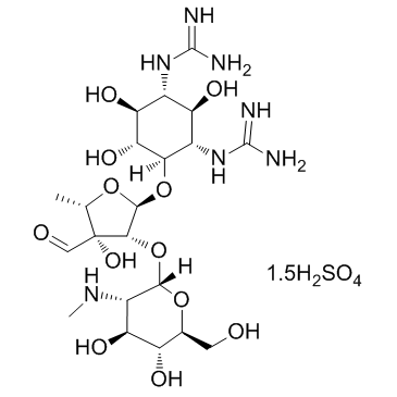
Steptomycin sulfate Structure Steptomycin sulfate
CAS:3810-74-0
acetic acid Structure acetic acid
CAS:1173022-32-6
acetic acid Structure acetic acid
CAS:64-19-7
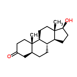
Stanolone Structure Stanolone
CAS:521-18-6