| In Vitro |
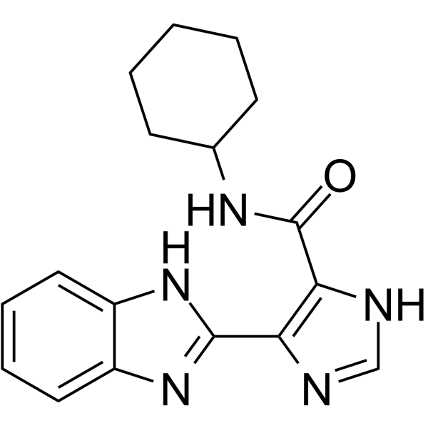
Autophagy-IN-2 (Compound 7h) (0-200 μM, 48 h) shows anti-viability activities against various cancer cells[1]. Autophagy-IN-2 (0-20 μM, 0-48 h) suppresses autophagic flux and impairs DNA repair through down-regulating chromatin ubiquitination in a p62-dependent manner[1]. Autophagy-IN-2 (0-20 μM, 48 h) induces cell cycle arrest at S-phase and mitochondria-dependent intrinsic apoptosis[1]. Cell Viability Assay[1] Cell Line: MDA-MB-231, MDA-MB-468, SCC25, HSC-3, HCT116, SW620, PC3, Aspc, PNAC, U87, U251 and LN229 Concentration: 0-200 μM Incubation Time: 48 h Result: Showed anti-viability activities with IC50 values of 8.31 ± 1.05, 6.03 ± 0.82, 18.64 ± 2.92, 24.03 ± 3.75, 35.53 ± 7.52, 25.43 ± 2.42, 17.48 ± 1.27, 26.89 ± 5.23, 19.25 ± 3.96, 15.37 ± 1.78, 12.92 ± 2.38 and 61.35 ± 11.61 μM against MDA-MB-231, MDA-MB-468, SCC25, HSC-3, HCT116, SW620, PC3, Aspc, PNAC, U87, U251 and LN229 cells, respectively. Western Blot Analysis[1] Cell Line: MDA-MB-231 and MDA-MB-468 Concentration: 5, 10 and 20 μM Incubation Time: 0, 6, 12, 24, 36 and 48 h Result: Significantly induced to LC3B-II in a dose - and time-dependent manner, resulted in a p62 accumulation in TNBC cells, increased γH2AX in a dose and time-dependent manner, activated the phosphorylation of ATM, NBS1 and SMC1, increased p62 in the nucleus and cytoplasm in a dose-dependent manner, and significantly reduced DNA-damage-induced formation of H2A poly-ubiquitin chains. Decreased the cellular levels of Cyclin A, Cyclin B, CDK1 and CDK2, increased P21 and the phosphorylation levels of CHK1 (Serine 345) and CHK2 (Threonine 68), and increased cytochrome c, cleaved caspase-3, cleaved caspase-9 and cleaved PAPR in a dose-dependent manner. Cell Cycle Analysis[1] Cell Line: MDA-MB-231 and MDA-MB-468 Concentration: 5, 10 and 20 μM Incubation Time: 48 h Result: Induced cell cycle arrest at S-phase. Apoptosis Analysis[1] Cell Line: MDA-MB-231 and MDA-MB-468 Concentration: 5, 10 and 20 μM Incubation Time: 48 h Result: Dramatically induced cell apoptosis in TNBC cells and obviously increase the proportion of late-phase apoptosis in a dose-dependent manner.
|