| Description |
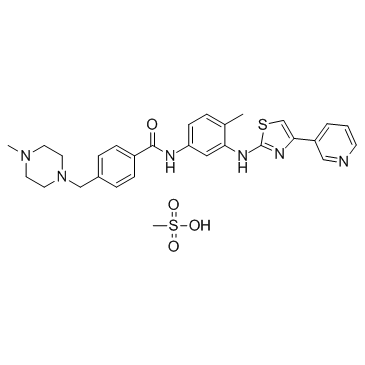
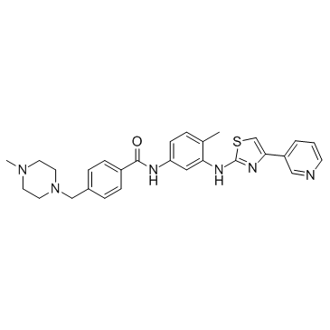
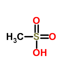
Masitinib mesylate is a novel inhibitor for Kit and PDGFRα/β with IC50 of 200 nM and 540 nM/800 nM, and has weak inhibition to ABL and c-Fms.
|
| Related Catalog |
|
| Target |
IC50: 200 nM (Kit), 540 nM (PDGFRα), 800 nM (PDGFRβ)
|
| In Vitro |
Masitinib is a competitive inhibitor against ATP at concentrations ≤500 nM. Masitinib also potently inhibits recombinant PDGFR and the intracellular kinase Lyn, and to a lesser extent, fibroblast growth factor receptor 3. In contrast, masitinib demonstrates weak inhibition of Abl and c-Fms. Masitinib more strongly inhibits degranulation, cytokine production, and bone marrow mast cell migration than imatinib. In Ba/F3 cells expressing human wild-type Kit, masitinib inhibits SCF (stem cell factor)-induced cell proliferation with an IC50 of 150 nM, while the IC50 for inhibition of IL-3-stimulated proliferation is at approximately >10 µM. In Ba/F3 cells expressing PDGFRα, masitinib inhibits PDGF-BB-stimulated proliferation and PDGFRα tyrosine phosphorylation with IC50 of 300 nM. Masitinib also causes inhibition of SCF-stimulated tyrosine phosphorylation of human Kit in mastocytoma cell-lines and BMMC. Masitinib inhibits Kit gain-of-function mutants, including V559D mutant and Δ27 mouse mutant with IC50 of 3 and 5 nM in Ba/F3 cells. Masitinib inhibits the cell proliferation of mastocytoma cell lines including HMC-1α155 and FMA3 with IC50 of 10 and 30 nM, respectively[1]. Masitinib inhibits cell growth and PDGFR phosphorylation in two novel ISS cell lines, which suggest that Masitinib displays activity against both primary and metastatic ISS cell line and may aid in the clinical management of ISS[2].
|
| In Vivo |
Masitinib inhibits tumour growth and increases the median survival time in Δ27-expressing Ba/F3 tumor models at 30 mg/kg, without cardiotoxicity or genotoxicity[1]. Masitinib (12.5 mg/kg/d, p.o.) increases overall TTP (time-to-tumor progression) compared with placebo in dogs[3]. The combination of masitinib/gemcitabine shows synergy in vitro on proliferation of gemcitabine-refractory cell lines Mia Paca2 and Panc1, and to a lesser extent on Mia Paca-2 pancreatic tumours in mice[4].
|
| Kinase Assay |
A 96-well microtitre plateis coated overnight with 0.25 mg/mL poly(Glu,Tyr 4:1), rinsed twice with 250 µL of washing buffer (10 mM phosphate-buffered saline [pH 7.4] and 0.05% Tween 20) and dried for 2 hours at room temperature. Assays are performed at room temperature with a final volume of 50 µL in kinase buffer (10 mM MgCl2, 1 mM MnCl2, 1 mM sodium orthovanadate, 20 mM HEPES, pH 7.8) containing ATP at a concentration of at least twice the Km for each enzyme and an appropriate amount of recombinant enzyme to ensure a linear reaction rate. Reactions are initiated upon introduction of the enzyme and terminated with the addition of one reaction volume (50 μL) of 100 mM EDTA per 5mol/Lurea mix. Plates are washed three times and incubated with 1:30,000 horseradish peroxidase-conjugated anti-phosphotyrosine monoclonal antibody, then washed three times and incubated with tetramethylbenzidine. The final reaction product is quantified by spectrophotometry at 450 nm.
|
| Cell Assay |
For the assay of Ba/F3 cell proliferation, microtitre plates are seeded with a total of 104 cells/well in 100 μL of RPMI 1640 medium with 10% foetal bovine serum at 37°C. These are supplemented, or not, with either 0.1% conditioned medium from X63-IL-3 cells or 250 ng/mL murine SCF. The murine SCF, which activates Kit, is purified from the conditioned medium of SCF-producing CHO cells. Cells are grown for 48 hours at 37°C with masitinib and then incubated with 10 μL/well of WST-1 reagent for 3 hours at 37°C. The amount of formazan dye formed is quantified by its absorbance at 450 nm using a scanning multiwell spectrophotometer. A blank well without cells is used as a background control for the spectrophotometer.
|
| Animal Admin |
Male Nog-SCID mice (7 weeks old) are under specific pathogen-free conditions at 20±1°C in a 12-hour light/12-hour dark cycle and ad libitum access to food and filtered water. Mia Paca-2 cells are cultured as described above. At day 0 (D0), mice are injected with 107 Mia Paca-2 cells in 200 µL PBS into the right flank. Tumours are allowed to grow for 1.5 to 4 weeks until the desired tumour size is reached (appr 200 mm3). At day 28, animals are allocated into four treatment groups (n=7 to 8 per group), ensuring that each group's mean body weight and tumour volume are well matched. Treatment is then administered for up to 4 weeks, after which time the animals are sacrificed. Treatments consisted of either: a) daily sterile water for the control group, b) an intraperitoneal (i.p.) injection of 50 mg/kg gemcitabine twice a week, c) daily gavage with 100 mg/kg masitinib, or d) combined i.p injection of 50 mg/kg gemcitabine twice a week and daily gavage with 100 mg/kg masitinib. Tumour size is measured with callipers and tumour volume is estimated using the formula: volume=(length×width2)/2. The tumour growth inhibition ratio is calculated as (100)×(median tumour volume of treated group)/(median tumour volume of control group).
|
| References |
[1]. Dubreuil P, et al. Masitinib (AB1010), a Potent and Selective Tyrosine Kinase Inhibitor Targeting KIT. PLoS One, 2009, 4(9), e7258. [2]. Lawrence J, et al. Masitinib demonstrates anti-proliferative and pro-apoptotic activity in primary and metastatic feline injection-site sarcoma cells. Vet Comp Oncol, 2011, doi: 10.1111/j.1476-5829.2011.00291.x. [3]. Hahn KA, et al. Masitinib is safe and effective for the treatment of canine mast cell tumors. J Vet Intern Med, 2008, 22(6), 1301-1309. [4]. Humbert M, et al. Masitinib combined with standard gemcitabine chemotherapy: in vitro and in vivo studies in human pancreatic tumour cell lines and ectopic mouse model. PLoS One. 2010 Mar 4;5(3):e9430.
|