1252003-15-8
1252003-15-8 structure
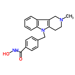
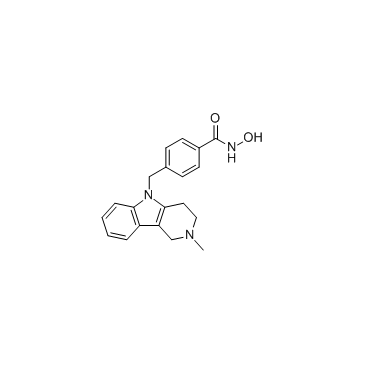
- Name: Tubastatin A
- Chemical Name: N-Hydroxy-4-[(2-methyl-1,2,3,4-tetrahydro-5H-pyrido[4,3-b]indol-5 -yl)methyl]benzamide
- CAS Number: 1252003-15-8
- Molecular Formula: C20H21N3O2
- Molecular Weight: 371.861
- Catalog: Biochemical Inhibitor Epigenetics HDAC inhibitor
- Create Date: 2018-12-19 21:43:30
- Modify Date: 2026-07-02 19:18:50
-
Tubastatin A is a potent and selective HDAC6 inhibitor with IC50 of 15 nM in a cell-free assay, and is selective (1000-fold more) against all other isozymes except HDAC8 (57-fold more).
| Name | N-Hydroxy-4-[(2-methyl-1,2,3,4-tetrahydro-5H-pyrido[4,3-b]indol-5 -yl)methyl]benzamide |
|---|---|
| Synonyms |
N-Hydroxy-4-[(2-methyl-1,2,3,4-tetrahydro-5H-pyrido[4,3-b]indol-5-yl)methyl]benzamide hydrochloride (1:1)
Tubastatin-A Tubastatin A Benzamide, N-hydroxy-4-[(1,2,3,4-tetrahydro-2-methyl-5H-pyrido[4,3-b]indol-5-yl)methyl]-, hydrochloride (1:1) TSLDASIIWAMMQN N-hydroxy-4-(2-methyl-1,2,3,4-tetrahydro-pyrido[4,3-b]indol-5-ylmethyl)benzamide |
| Description | Tubastatin A is a potent and selective HDAC6 inhibitor with IC50 of 15 nM in a cell-free assay, and is selective (1000-fold more) against all other isozymes except HDAC8 (57-fold more). |
|---|---|
| Related Catalog | |
| Target |
HDAC6:15 nM (IC50) HDAC8:854 nM (IC50) HDAC1:16400 nM (IC50) |
| In Vitro | Tubastatin A is substantially selective for all 11 HDAC isoforms and maintains over 1000-fold selectivity against all isoforms excluding HDAC8, where it has approximately 57-fold selectivity. In homocysteic acid (HCA) induced neurodegeneration assays, Tubastatin A displays dose-dependent protection against HCA-induced neuronal cell death starting at 5 μM with near complete protection at 10 μM[1]. At 100 ng/mL Tubastatin A increases Foxp3+ T-regulatory cells (Tregs) suppression of T cell proliferation in vitro[2]. Tubastatin A treatment in CC12 cells would lead to myotube formation impairment when alpha-tubulin is hyperacetylated early in the myogenic process; however, myotube elongation occurs when alpha-tubulin is hyeperacetylated in myotubes[3]. A recent study indicates that Tubastatin A treatment increases cell elasticity as revealed by atomic force microscopy (AFM) tests without exerting drastic changes to the actin microfilament or microtubule networks in mouse ovarian cancer cell lines, MOSE-E and MOSE-L[4]. |
| In Vivo | Daily treatment of Tubastatin A at 0.5 mg/kg inhibits HDAC6 to promote Tregs suppressive activity in mouse models of inflammation and autoimmunity, including multiple forms of experimental colitis and fully major histocompatibility complex (MHC)-incompatible cardiac allograft rejection[2]. |
| Kinase Assay | Enzyme inhibition assays are performed using the Reaction Biology HDAC Spectrum platform. The HDAC1, 2, 4, 5, 6, 7, 8, 9, 10, and 11 assays use isolated recombinant human protein; HDAC3/NcoR2 complex is used for the HDAC3 assay. Substrate for HDAC1, 2, 3, 6, 10, and 11 assays is a fluorogenic peptide from p53 residues 379-382 (RHKKAc); substrate for HDAC8 is fluorogenic diacyl peptide based on residues 379-382 of p53 (RHKAcKAc). Acetyl-Lys (trifluoroacetyl)-AMC substrate is used for HDAC4, 5, 7, and 9 assays. Tubastatin A is dissolved in DMSO and tested in 10-dose IC50 mode with 3-fold serial dilution starting at 30 μM. Control Compound Trichostatin A (TSA) is tested in a 10-dose IC50 with 3-fold serial dilution starting at 5 μM. IC50 values are extracted by curve-fitting the dose/response slopes. |
| Cell Assay | Primary cortical neuron cultures are obtained from the cerebral cortex of fetal Sprague-Dawley rats (embryonic day 17) as described previously. All experiments are initiated 24 hours after plating. Under these conditions, the cells are not susceptible to glutamate-mediated excitotoxicity. For cytotoxicity studies, cells are rinsed with warm PBS and then placed in minimum essential medium containing 5.5 g/L glucose, 10% fetal calf serum, 2 mM L-glutamine, and 100 μM cystine. Oxidative stress is induced by the addition of the glutamate analogue homocysteate (HCA; 5 mM) to the media. HCA is diluted from 100-fold concentrated solutions that are adjusted to pH 7.5. In combination with HCA, neurons are treated with Tubastatin A at the indicated concentrations. Viability is assessed after 24 hours by MTT assay (3-(4,5-dimethylthiazol-2-yl)-2,5-diphenyltetrazolium bromide) method. |
| Animal Admin | The effects of HDAC6 targeting in dextran sodium sulfate (DSS) and adoptive transfer models of colitis are evaluated, using 10 mice per group. Freshly prepared 4% (wt/vol) DSS (MP Biomedicals) is added daily for 5 days to the pH-balanced tap water of WT B6 mice. Mice are treated daily for 7 days with tubacin or niltubacin (0.5 mg/kg of body weight/day, i.p.), and colitis is assessed by daily monitoring of body weight, stool consistency, and fecal blood. Stool consistency is scored as 0 (hard), 2 (soft), or 4 (diarrhea), and fecal blood (Hemoccult) is scored as 0 (absent), 2 (occult), or 4 (gross). To assess prevention of colitis in a T cell-dependent model, CD4+ CD45RBhi T cells (1×10⁶) isolated from WT mice using magnetic beads (>95% cell purity, flow cytometry) are injected i.p. into B6/Rag1−/− mice plus CD4+ CD25+ Tregs (1.25×10⁵) isolated using magnetic beads from HDAC6−/− or WT mice (>90% Treg purity, flow cytometry) and mice are monitored biweekly for clinical evidence of colitis. To assess therapy of established T cell-dependent colitis, B6/Rag1−/− mice are injected i.p. with CD4+ CD45RBhi cells (1×10⁶). Once colitis has developed, mice also receive CD4+ CD25+ Tregs (5×10⁵ cells) isolated as described above from HDAC6−/− or WT mice or treatment with HDAC6i (tubastatin A) or HSP90i (17-AAG). Mice are monitored for continued weight loss and stool consistency. At the cessation of the study, paraffin sections of colons stained with Alcian Blue or hematoxylin and eosin are graded histologically or evaluated by immunoperoxidase staining for Foxp3+ Treg infiltration. |
| References |
| Molecular Formula | C20H21N3O2 |
|---|---|
| Molecular Weight | 371.861 |
| Exact Mass | 371.140045 |
| PSA | 57.50000 |
| LogP | 3.12530 |
| Storage condition | 2~8°C |
| Hazard Codes | Xi |
|---|
| Precursor 0 | |
|---|---|
| DownStream 1 | |