| In Vitro |
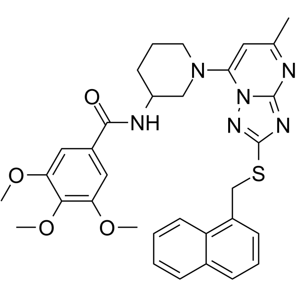
Antitumor agent-55 (compound 5q) shows inhibitory activity against MCF-7, PC3, MGC-803, PC9, and WPMY-1 (normal human prostatic stromal myofibroblast cell line), with IC50 values of 11.54 ± 0.18, 0.91 ± 0.31, 8.21 ± 0.50, 34.68 ± 0.67, and 48.15 ± 0.33, respectively[1]. Antitumor agent-55 (0-10 μM, 24-72 h) significantly inhibits the proliferation of PC3 cells dose- and time-dependently[1]. Antitumor agent-55 (0-4 μM, 24 h) increases the G1/S phase population, and dose-dependently elevates the expression of p27 protein[1]. Antitumor agent-55 (0-4 μM, 24-48 h) dose-dependently induces the accumulation of ROS, and induces apoptosis of PC3 cells through activating the two apoptotic signaling pathways simultaneously[1]. Antitumor agent-55 (0-1 μM, 48 h) effectively inhibits the wound healing and the migration of PC3 cells in a dose-dependent manner[1]. Cell Viability Assay Cell Line: PC3 cells[1] Concentration: 0, 0.156, 0.313, 0.625, 1.25, 2.5, 5, 10 μM Incubation Time: 24, 48, 72 h Result: Significantly inhibited the proliferation of PC3 cells dose- and time-dependently, formed fewer and smaller colonies. Cell Cycle Analysis Cell Line: PC3 cells[1] Concentration: 0, 1, 2, 4 μM Incubation Time: 24 h Result: Significantly increased the G1/S phase population while decreased G2/M content at high concentration in PC3 cells. Western Blot Analysis Cell Line: PC3 cells[1] Concentration: 0, 1, 2, 4 μM Incubation Time: 24 h, 48 h Result: Dose-dependently elevated the expression of p27 protein, markedly elevated the expression of pro-apoptotic Bax and P53 while anti-apoptotic Bcl-2 expression was down-regulated, and significantly increased the expression of cleaved caspase 3/9 and cleaved PARP in a dose-dependent manner. Apoptosis Analysis Cell Line: PC3 cells[1] Concentration: 0, 1, 2, 4 μM Incubation Time: 48 h Result: Dose-dependently led to significant increase of apoptotic population, and the apoptotic percentage was up to 70.7% at 4 μM, which was far higher than the control group (3.5%).
|
