| Structure | Name/CAS No. | Articles |
|---|---|---|
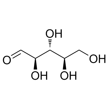 |
D-Ribose
CAS:50-69-1 |
|
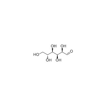 |
D-(+)-Glucose
CAS:50-99-7 |
|
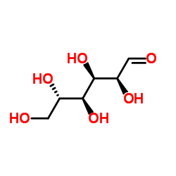 |
L-Glucose
CAS:921-60-8 |
|
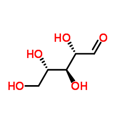 |
L-(+)-Ribose
CAS:147-81-9 |