| Structure | Name/CAS No. | Articles |
|---|---|---|
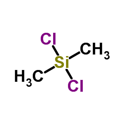 |
Dichlorodimethylsilane
CAS:75-78-5 |
|
 |
3-(2-Aminoethylamino)propyltrimethoxysilane
CAS:1760-24-3 |