| Structure | Name/CAS No. | Articles |
|---|---|---|
 |
sodium chloride
CAS:7647-14-5 |
|
 |
Forskolin
CAS:66575-29-9 |
|
 |
SodiuM bicarbonate
CAS:144-55-8 |
|
 |
Trypsin
CAS:9002-07-7 |
|
 |
HEPES
CAS:7365-45-9 |
|
 |
SODIUM CHLORIDE-35 CL
CAS:20510-55-8 |
|
 |
Cyclothiazide
CAS:2259-96-3 |
|
 |
Poly-D-lysine hydrobromide (MW 30000-70000)
CAS:27964-99-4 |
|
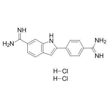 |
4',6-Diamidino-2-phenylindole dihydrochloride
CAS:28718-90-3 |
|
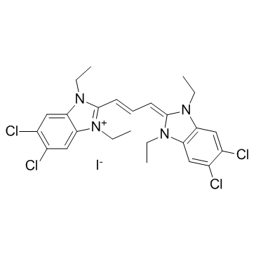 |
JC-1
CAS:3520-43-2 |