| Structure | Name/CAS No. | Articles |
|---|---|---|
 |
sodium chloride
CAS:7647-14-5 |
|
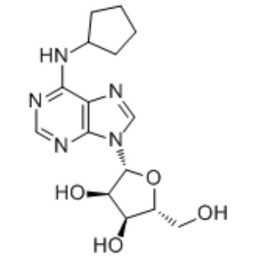 |
N6-Cyclopentyladenosine
CAS:41552-82-3 |
|
 |
SODIUM CHLORIDE-35 CL
CAS:20510-55-8 |
|
 |
γ-Linolenoyl-CoA
CAS:18172-33-3 |
|
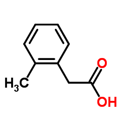 |
o-Tolylacetic acid
CAS:644-36-0 |
|
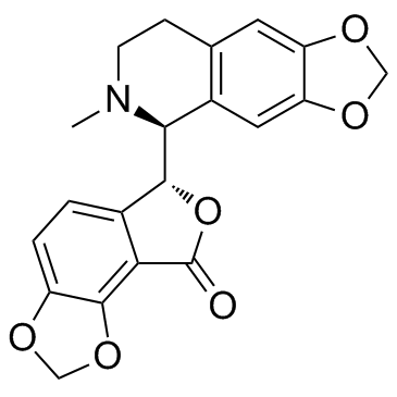 |
(+)-Bicuculline
CAS:485-49-4 |