| Description |
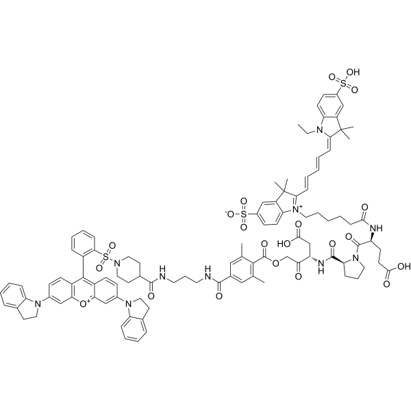
LE 28 is a selective and activity-dependent legumain probe. LE 28 becomes fluorescent only upon binding active legumain. LE 28 can be used for research of cancers and inflammatory diseases[1].
|
| Related Catalog |
|
| In Vitro |
LE 28 (0-5 μM, 1 h) 可以通过裂解和荧光 SDS-PAGE 标记法检测 RAW 细胞裂解物中的 legumain[1]。 LE 28 (1 μM, 5 h) 在 RAW 细胞中与 Lysotracker 共定位较好[1]。
|
| In Vivo |
LE 28 (2 mg/kg, i.v.) 在小鼠肾脏中产生特异性信号[1]。 LE 28 (2 mg/kg, i.v.) 在 HCT-116 异种移植肿瘤模型中 30 分钟时在肿瘤周围检测到信号,7 小时后肿瘤与正常组织之间的信噪比达到最大[1]。 Animal Model: HCT-116 xenograft tumor model[1] Dosage: 2 mg/kg Administration: i.v. Result: Detected the signal around the periphery of the tumors in as early as 30 min, with maximal contrast between tumor and normal tissue achieved after seven hours, and the signal remained constant up to 28 h.
|
| References |
[1]. Edgington LE, et al. Functional imaging of legumain in cancer using a new quenched activity-based probe. J Am Chem Soc. 2013 Jan 9;135(1):174-82.
|
