cypate
Modify Date: 2025-08-26 16:59:00
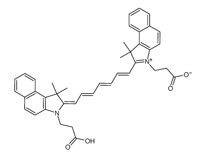
cypate structure
|
Common Name | cypate | ||
|---|---|---|---|---|
| CAS Number | 95837-47-1 | Molecular Weight | 624.76700 | |
| Density | N/A | Boiling Point | N/A | |
| Molecular Formula | C41H40N2O4 | Melting Point | N/A | |
| MSDS | N/A | Flash Point | N/A | |
Use of cypateCypate, a cyanine dye, is a near infrared (NIR) fluorescent probe for in vivo tumor imaging[1][2]. |
| Name | cypate |
|---|
| Description | Cypate, a cyanine dye, is a near infrared (NIR) fluorescent probe for in vivo tumor imaging[1][2]. |
|---|---|
| Related Catalog | |
| In Vitro | Guidelines (Following is our recommended protocol. This protocol only provides a guideline, and should be modified according to your specific needs). 1. 将每种类型细胞在单独的 35 毫米玻璃底培养皿上接种 1×10⁶ 个。 2. 24 小时后,将细胞与 10 µM Cypate 无酚红培养基溶液一起孵育 2 小时。 3. 处理后,细胞用 PBS 洗涤 3 次并用 4% 多聚甲醛固定用于成像,或收集细胞内的 Cypate 内容物用于光谱和/或 LC-MS 分析。 4. 对体外细胞培养实验进行共聚焦显微镜检查。Cypate 的激发波长为 647 nm[1]。 |
| In Vivo | Cypate (10 nmol; 100 µL; 静脉给药; 每 24 小时一次; 连续 6 天) 导致肝脏蓄积最高[1]。 Cypate (5 mg/kg; 静脉给药) 在具有 4T1 细胞的 Balb/c 小鼠肿瘤中的荧光信号微弱并迅速衰减,这可能是由于游离 Cypate 从体内快速消除所致。在游离cypate组中观察到肝脏和肾脏中更强的荧光。使用 IVIS Lumina 成像系统 (Ex-745 nm; Em-800 nm) 评估 Cypate 荧光信号[2]。 Animal Model: Foxn1nu/Foxn1nu nude 2.5-month-old female mice with breast cancer cells (MDA-MB-231 Luc2)[1] Dosage: 10 nmol in 100 µL PBS Administration: IV; every 24 hours for 6 days Result: The accumulation alone in the tumor was negligible for 24 hours after the injection. The liver had the highest accumulation at all time points. |
| References |
| Molecular Formula | C41H40N2O4 |
|---|---|
| Molecular Weight | 624.76700 |
| Exact Mass | 624.29900 |
| PSA | 83.68000 |
| LogP | 7.35800 |