IWP-2
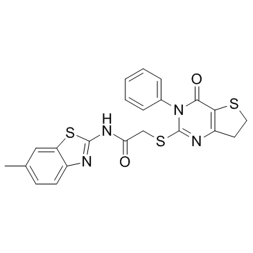
IWP-2 structure
|
Common Name | IWP-2 | ||
|---|---|---|---|---|
| CAS Number | 686770-61-6 | Molecular Weight | 466.599 | |
| Density | 1.5±0.1 g/cm3 | Boiling Point | N/A | |
| Molecular Formula | C22H18N4O2S3 | Melting Point | 257 °C(dec.) | |
| MSDS | USA | Flash Point | N/A | |
Use of IWP-2IWP-2 is an inhibitor of Wnt processing and secretion with IC50 of 27 nM. |
| Name | N-(6-Methyl-2-benzothiazolyl)-2-[(3,4,6,7-tetrahydro-4-oxo-3-phenylthieno[3,2-d]pyrimidin-2-yl)thio]-acetamide |
|---|---|
| Synonym | More Synonyms |
| Description | IWP-2 is an inhibitor of Wnt processing and secretion with IC50 of 27 nM. |
|---|---|
| Related Catalog | |
| Target |
IC50: 27 nM (Wnt)[1] |
| In Vitro | IWP-2, an inhibitor of WNT processing and secretion. IWP-2 significantly enhances the anti-proliferative effect of LEF. It is also obvious that the combination of LEF and IWP-2 could minimize the expression of β-catenin, c-Myc, Cyclin D1, Bcl2 and Bax to the largest extent compared with single agents[2]. Following treatment in the MKN28 cell line for four days, 10-50 μM IWP-2 significantly suppressed the proliferation of MKN28 cells (P<0.05). In addition, anchor-dependent and anchor-independent colony numbers are significantly decreased following IWP-2 treatment (P<0.05)[3]. |
| In Vivo | To evaluate the efficacy of IWP-2 in vivo, 200 μL each of IWP-2-liposome or free liposome i separately injected into C57BL/6 mice intraperitoneally about 2 h before injection of a similar volume of either blue-dye-filled latex beads or E. coli DH5α. IWP-2 causes significant reduction in the uptake of blue beads as well as E. coli as assessed by CFUs in peritoneal lavage cells within 2 h. In addition, the levels of TNF-α and IL-6 in the lavage fluid of the corresponding mice are reduced by 2-4-fold compared with control values. Interestingly, IWP-2 even induces a considerable increase in secretion of the anti-inflammatory cytokine IL-10[4]. Pretreatment with IWP-2 significantly (P<0.05) abolished SP-induced increase of Wnt3a, p-GSK3β, and β-catenin expressions[5]. |
| Cell Assay | The human RCC cell lines 786O and Caki-2 (5×10³) are seeded into 96-well plates. Cell viability is estimated by MST assay after Caki-2 acells are incubated with ncreasing concentrations of LEF together with 20 μM IWP-2 for 48 h.After treatment, 10 μL MTS is added into each well for 2 h incubation. The absorbance is measured using a model ELX800 Micro Plate Reader at 490 nm. For colony formation assay, Caki-2 cells are trypsinized to single cell suspensions and seeded into fresh 6-well plates at 1000 cells/well. Then cells are incubated with LEF at depicted concentrations for 7 days. Colonies are fixed with absolute methanol for 15 min and then stained with 0.1% crystal violet for 20 min. After washing with PBS three times, the colonies with a diameter over 2 mm are visualized by a digital camera[2]. |
| Animal Admin | Mice[4] About 3-mo-old C57BL/6 mice are housed four to five in a cage at 23°C in a 12-h light/dark cycle. Mice are injected intraperitoneally (i.p.) first with either 200 μL of liposome-IWP2 (LI) or liposome (L) and then after 2 h with 1×10⁸ or 2×10⁸ CFU E. coli in 200 μL of sterile PBS. After 2 h or 24 h mice are killed, and the peritoneal cavity is washed with 5 mL of sterile ice-cold PBS. The peritoneal lavage fluid is centrifuged at 300× g for 5 min, the cell pellet is resuspended in RPMI 1640 complete medium, and the supernatant is used for cytokine assay. For ex vivo experiments, peritoneal phagocytes are isolated as above from normal mice, and equal numbers of cells are plated in medium overnight at 37°C in 5% CO2 before performing further experiments. Rats[5] Adult, male, and healthy Wistar rats weighing 220-280 g are used. Rats are randomly divided into 6 groups as follows (n=72, 12 per group): (1) Sham group (Group S), (2) I/R group (Group I/R), (3) I/R+DMSO group (Group DMSO), (4) I/R+IWP group (Group IWP), (5) SP group (Group SP), and (6) SP+Wnt inhibitor IWP-2 group (Group SP+IWP). The hearts are continuously perfused for 120 min in Group S. After 10 min of equilibration, the isolated hearts are continuously perfused for 20 min, then subjected to 30 min of ischemia followed by 60 min of reperfusion in Group I/R; Groups DMSO, IWP, SP and SP+IWP receive 15 min of perfusion with K-H solution containing 0.5 mL/L DMSO, 10 µM IWP (SIGMA-ALDRICH, USA), 2.4 vol% Sevoflurane, 2.4 vol% Sevoflurane+10 µM IWP, respectively, followed by 5 min washout before I/R. |
| References |
| Density | 1.5±0.1 g/cm3 |
|---|---|
| Melting Point | 257 °C(dec.) |
| Molecular Formula | C22H18N4O2S3 |
| Molecular Weight | 466.599 |
| Exact Mass | 466.059174 |
| PSA | 156.11000 |
| LogP | 5.25 |
| Index of Refraction | 1.787 |
| InChIKey | WRKPZSMRWPJJDH-UHFFFAOYSA-N |
| SMILES | Cc1ccc2nc(NC(=O)CSc3nc4c(c(=O)n3-c3ccccc3)SCC4)sc2c1 |
| Storage condition | Store at +4°C |
| Water Solubility | DMSO: >5mg/mL |
| RIDADR | NONH for all modes of transport |
|---|
|
Ligand-independent canonical Wnt activity in canine mammary tumor cell lines associated with aberrant LEF1 expression.
PLoS ONE 9(6) , e98698, (2014) Pet dogs very frequently develop spontaneous mammary tumors and have been suggested as a good model organism for breast cancer research. In order to obtain an insight into underlying signaling mechani... |
|
|
Notch inhibition induces mitotically generated hair cells in mammalian cochleae via activating the Wnt pathway.
Proc. Natl. Acad. Sci. U. S. A. 112(1) , 166-71, (2015) The activation of cochlear progenitor cells is a promising approach for hair cell (HC) regeneration and hearing recovery. The mechanisms underlying the initiation of proliferation of postnatal cochlea... |
|
|
A switch from canonical to noncanonical Wnt signaling mediates early differentiation of human neural stem cells.
Stem Cells 32(12) , 3196-208, (2014) Wnt/β-catenin signaling is essential for neurogenesis but less is known about β-catenin-independent Wnt signals. We show here that Wnt/activator protein-1 (AP-1) signaling drives differentiation of hu... |
| N-(6-Methyl-1,3-benzothiazol-2-yl)-2-[(4-oxo-3-phenyl-3,4,6,7-tetrahydrothieno[3,2-d]pyrimidin-2-yl)sulfanyl]acetamide |
| Acetamide, N-(6-methyl-2-benzothiazolyl)-2-[(3,4,6,7-tetrahydro-4-oxo-3-phenylthieno[3,2-d]pyrimidin-2-yl)thio]- |
| IWP-2 |