173897-44-4
173897-44-4 structure
- Name: Y-39983 HCl
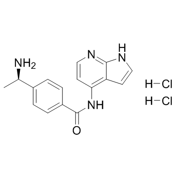
- Chemical Name: 4-[(1R)-1-aminoethyl]-N-(1H-pyrrolo[2,3-b]pyridin-4-yl)benzamide,dihydrochloride
- CAS Number: 173897-44-4
- Molecular Formula: C16H18Cl2N4O
- Molecular Weight: 353.24600
- Catalog: Signaling Pathways Cell Cycle/DNA Damage ROCK
- Create Date: 2016-05-18 04:43:11
- Modify Date: 2026-07-29 06:47:56
-
Y-33075 dihydrochloride is a selective ROCK inhibitor derived from Y-27632, and is more potent than Y-27632, with an IC50 of 3.6 nM.
| Name | 4-[(1R)-1-aminoethyl]-N-(1H-pyrrolo[2,3-b]pyridin-4-yl)benzamide,dihydrochloride |
|---|---|
| Synonyms |
(R)-4-(1-Aminoethyl)-N-(1H-pyrrolo[2,3-b]pyridin-4-yl)benzamide dihydrochloride
CS-0096 Y-33075 (dihydrochloride) |
| Description | Y-33075 dihydrochloride is a selective ROCK inhibitor derived from Y-27632, and is more potent than Y-27632, with an IC50 of 3.6 nM. |
|---|---|
| Related Catalog | |
| Target |
ROCK:3.6 nM (IC50) PKC:420 nM (IC50) CaMKII:810 nM (IC50) |
| In Vitro | Y-33075 (Y-39983) is a potent ROCK inhibitor, with an IC50 of 3.6 nM. Y-33075 also inhibits PKC and CaMKII more potently than Y-27632, and the IC50s of Y-27632 and Y-33075 for PKC are 9.0 μM and 0.42 μM, respectively, whereas the IC50s of Y-27632 and Y-33075 for CaMKII are 26 μM and 0.81 μM, respectively. The IC50s of Y-27632 and Y-33075 for PKC is 82 and 117 times those for ROCK, respectively, whereas the IC50s of Y-27632 and Y-33075 for CaMKII is 236 and 225 times those for ROCK, respectively[1]. Y-33075 (Y-39983, 10 μM) extends neurites in the retinal ganglion cells (RGCs) compared with those in RGCs treated without Y-39983[2]. Y-33075 (Y-39983, 1 μM) inhibits the contraction of rabbit ciliary artery segments evoked by histamine in Ca2+-free solutions. Y-33075 (10 μM) shows no effect on the [Ca2+]i increase with the high-potassium (high-K) solution[3]. |
| In Vivo | In rabbits, Y-39983 (≥0.01%) significantly lowers intraocular pressure (IOP) at 2 hours after topical administration. In monkeys, Y-39983 (0.05%)-treated eyes show significant reduction of IOP between 2 and 7 hours after topical administration[1]. Y-39983 (100 μM) increases the regenerating axons of retinal ganglion cells (RGCs) in the eyes of the rats[2]. |
| Kinase Assay | Recombinant ROCK (ROK α/ROCK II), purified protein kinase C (PKC: mixture of α, β, γ isoforms), and recombinant calmodulin-dependent protein kinase II (CaMK II) are used in the assay. ROCK (0.2 U/mL) is incubated with 1 μM [γ-32P] ATP and 10 μg/mL histone as substrates in the absence or presence of various concentrations of Y-27632, Y-33075, or staurosporine at room temperature for 20 minutes in 20 mM MOPS (3-(N-morpholino)propanesulfonic acid) buffer (pH 7.2) containing 0.1 mg/mL bovine serum albumin (BSA), 5 mM dithiothreitol [DTT], 10 mM β-glycerophosphate, 50 μM Na3VO4, and 10 mM MgCl2 in a total volume of 100 μL. PKC (10 ng/mL) is incubated with 1 μM [γ-32P] ATP and 20 μM PKC substrate in the absence or presence of various concentrations of Y-27632, Y-33075, or staurosporine at room temperature for 30 minutes in 20 mM MOPS buffer (pH 7.5) containing 0.1 mg/mL BSA, 10 mM DTT, 10 mM β-glycerophosphate, 50 μM Na3VO4, 2 mM CaCl2, 20 μg/mL phosphatidyl-l-serine, and 10 mM MgCl2 in a total volume of 100 μL. CaMK II (125 U/mL) is incubated with 1 μM [γ-32P] ATP, 10 μM calmodulin, and 20 μM CaMK II substrate, in the absence or presence of various concentrations of Y-27632, Y-33075, or staurosporine at room temperature for 30 minutes in 20 mM MOPS buffer (pH 7.5) containing 0.2 mg/mL BSA, 0.5 mM DTT, 0.1 mM β-glycerophosphate, 50 μM Na3VO4, 1 mM CaCl2, and 5 mM MgCl2 in a total volume of 100 μL. Incubation is terminated by the addition of 100 μL of 0.7% phosphoric acid. A 160 μL portion of the mixture is transferred to Multiscreen-PH plate. A positively charged phosphocellulose filter absorbs the substrate that binds 32P. The filter is washed with 300 μL of 0.5% phosphoric acid and then twice with purified water and then dried. The radioactivity of the dried filter is measured with a liquid scintillation counter. Results are presented as 50% inhibitory concentrations and 95% confidence intervals (CIs)[1]. |
| Cell Assay | In brief, retinal cell suspensions are obtained from dissected retinas of Wistar rats by papain treatment. Retinal ganglion cells (RGCs) are purified by the panning method using anti-rat CD11 antibody for removal of microglia cells and anti-rat Thy-1 antibody for isolation of ganglion cells. The purified RGCs (5000 cells/plate) are seeded into 24-well plates coated by 50 μg/mL of poly-l-lysine and 2 μg/mL of merosin, and are cultured in serum-free neurobasal medium supplemented with 2% B27 supplement, 50 ng/mL BDNF, 50 ng/mL CNTF, 5 μM forskolin, and 1 mM glutamine under a 95% air-5% CO2 atmosphere at 37°C. After completion of 24-hour cultivation, RGCs are cultured in medium with or without 10 μM Y-33075 as the final concentration for 24 hours and morphologically observed by phase-contrast microscopy. The concentration used is determined based on the effect of Y-33075 on trabecular meshwork contraction in vitro. Since this study is conducted in order to confirm whether Y-33075 has a potential of effect on axonal regeneration of RGCs, the effect is unquantitateively evaluated[2]. |
| Animal Admin | In brief, SD rats is anesthetized with an intraperitoneal injection of sodium pentobarbital (0.4 mg/kg body weight), and the optic nerve of one eye is transected 4 to 6 mm posterior to the eyeball, taking care to avoid injury to the ophthalmic artery. The anterior branch of the sciatic nerve is excised and sutured autologously to the optic nerve stump with nylon sutures. The other end of the graft is sutured to the temporalis muscle. A small piece (3 mm × 3 mm) of gelatin sponge soaked with 10 μM Y-33075 or saline as a control is implanted in the space behind the optic stump after optic nerve transection in intact animals. Five μL of 0.12 mM or 1.2 mM Y-33075 solution or saline is administered into the vitreous body to final concentrations of 10 μM or 100 μM, respectively. The concentrations of Y-33075 used is determined as 10 μM that is effective in the in vitro study on axonal regeneration of RGCs, and also as 100 μM in order to confirm the dose response of Y-33075. Six weeks after surgery, rats is anesthetized with an intraperitoneal injection of sodium pentobarbital (0.4 mg/kg body weight), and 4-Di-10ASP is embedded in the transplanted sciatic nerve to retrogradely label RGCs with axonal regeneration into the sciatic nerve. Three days after dye embedding, rats is euthanized and the eyes is enucleated for preparation of retinal flat-mounts. The posterior eyecup is then separated from the vitreous body and postfixed with 4% paraformaldehyde solution in phosphate buffer for around 1 hour at room temperature. Fluorescence micrographs of the labeled cells is imported using a fluorescence microscope connected to a computer. Labeled cells is counted using image analysis software. As a normal group, the subsequent procedure for retrograde labeling with 4-Di-10ASP is performed without grafting sciatic nerve and administering the test drug. Statistical analysis is performed using logarithmically transformed values due to differences in variance among the groups. The statistical significance of differences between the normal and saline groups and the saline and Y-33075 groups is examined by t-test (onesided) and William’s test (one-sided). Findings of p < 0.05 is considered significant[2]. |
| References |
| Molecular Formula | C16H18Cl2N4O |
|---|---|
| Molecular Weight | 353.24600 |
| Exact Mass | 352.08600 |
| PSA | 83.80000 |
| LogP | 5.21220 |
| Storage condition | -20℃ |