| In Vitro |
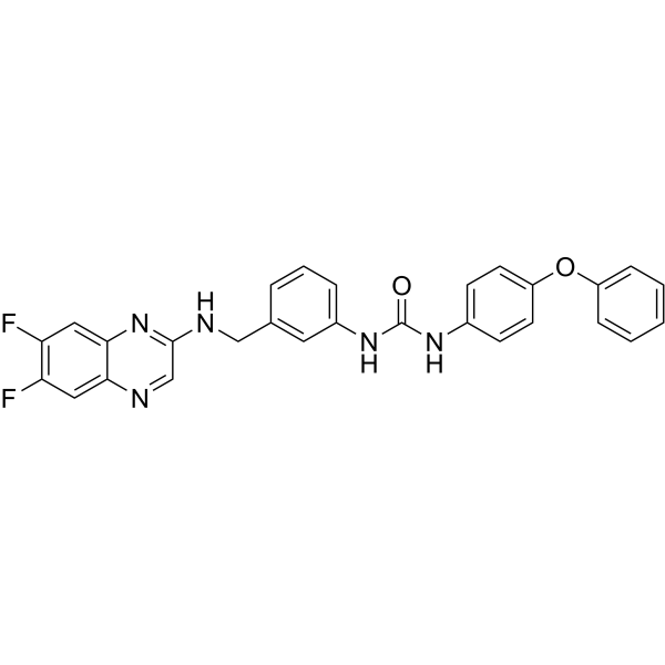
Anticancer agent 31 (compound 2d) shows anticancer activity against human tumor cell lines (MGC-803, NCI-H460, T-24, HeLa, HepG2, and SMMC-7721) and displays lower cytotoxicity than 5-FU (HY-90006), Sorafenib (HY-10201), and Cisplatin (HY-17394)[1]. Anticancer agent 31 (10, and 15 μM; 24 h) arrests cell cycle at S phase in MGC-803 cells, and induces tumor cells apoptosis[1]. Anticancer agent 31 (5, 10, and 15 μM; 24 h) reduces cell cycle regulatory protein CDK2, CDK4, cyclin A2, cyclin B1, and Apaf-1, anti-apoptotic protein Bcl-2; increases pro-apoptotic protein Bax protein level[1]. Anticancer agent 31 (5, 10 μM; 24 h) activates caspase-3 and caspase-9 by 73.6% and 65.3%, respectively; and also causes the loss of mitochondrial membrane potential (MMP) increase in JC-1 (HY-15534) detection[1]. Cell Cytotoxicity Assay[1] Cell Line: Normal cell line: HL-7702 and six human tumor cell lines: MGC-803, NCI-H460, T-24, HeLa, HepG2, and SMMC-7721 Concentration: Incubation Time: Result: Inhibited human tumor cell with IC50s of 9 μM (MGC-803), 12.3 μM (HeLa), 13.3 μM (NCI-H460), 30.4 μM (HepG2), 17.6 μM (SMMC-7721), 27.5 μM (T-24), respectively.Showed lowe cytotoxicity with an IC50 value of 80.9 μM, higher than the IC50s of tumor cell cells. Western Blot Analysis[1] Cell Line: MGC-803 Concentration: 0, 5, 10, 15 μM Incubation Time: 24 hours Result: Decreased cell cycle regulatory protein (cyclin-dependent kinase (CDK)2, CDK4, cyclin A2, and cyclin B1) levels in a dose-dependent manner. Suppressed anti-apoptotic protein Bcl-2 expression and up-regulated pro-apoptotic protein Bax level. Apoptosis Analysis[1] Cell Line: MGC-803 Concentration: 0, 5, 10, 15 μM Incubation Time: 24 hours Result: Caused MGC-803 cells arrest at S phase from 24.45% to 41.39% at 15 μM concentration. Indicated that there was a interaction with DNA in the nucleus and affect DNA replication. Increased the percentage of apoptotic tumor cells from 4.31% to 37.21%. Immunofluorescence[1] Cell Line: MGC-803 Concentration: 5, 10 μM Incubation Time: 24 hours; JC-1 as the fluorescent probe Result: Induced the loss of mitochondrial membrane potential (MMP) from 0.79% (control) to 37.4% (5 μM) and 81.4% (10 μM), respectively.
|
