- Shanghai Nianxing Industrial Co., Ltd
- China
- Product Name: Ginsenoside Rh3
- Price: Check
- Purity: 98.0%
- Stocking Period: 10 Day
- Contact: Yang
- Email: sales@echemcloud.com
- Dayang Chem (Hangzhou) Co., Ltd.
- China
- Product Name: Ginsenoside Rh3
- Price: $Inquiry/100g $Inquiry/1kg $Inquiry/100kg $Inquiry/1000kg
- Purity: 98.0%
- Stocking Period: Inquiry
- Contact: Ms Wang
- Email: enquiry@dycnchem.com
- Shanghai Jizhi Biochemical Technology Co., Ltd
- China
- Product Name: Ginsenoside Rh3
- Price: ¥3728.0/20mg
- Purity: 98.0%
- Stocking Period: 1 Day
- Contact: Liu jia


- Email: 2130147988@qq.com
- naturewill
- China
- Product Name: Ginsenoside Rh3
- Price: ¥Inquiry/5mg ¥Inquiry/20mg ¥Inquiry/100mg ¥Inquiry/1g
- Purity: 98.0%
- Stocking Period: Inquiry
- Contact: Bella
- Email: 3157321941@qq.com
105558-26-7
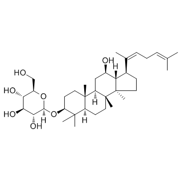
105558-26-7 structure
- Name: Ginsenoside Rh3
- Chemical Name: (2R,3S,4S,5R,6R)-2-(hydroxymethyl)-6-[[(8R,10R,12S,13R,14S,17S)-12-hydroxy-4,4,8,10,14-pentamethyl-17-[(2Z)-6-methylhepta-2,5-dien-2-yl]-2,3,5,6,7,9,11,12,13,15,16,17-dodecahydro-1H-cyclopenta[a]phenanthren-3-yl]oxy]oxane-3,4,5-triol
- CAS Number: 105558-26-7
- Molecular Formula: C36H60O7
- Molecular Weight: 604.857
- Catalog: Signaling Pathways NF-κB Keap1-Nrf2
- Create Date: 2018-06-08 17:05:50
- Modify Date: 2025-08-27 09:45:02
-
Ginsenoside Rh3 is a bacterial metabolite of Ginsenoside Rg5. Ginsenoside Rh3 treatment in human retinal cells induces Nrf2 activation.
| Name | (2R,3S,4S,5R,6R)-2-(hydroxymethyl)-6-[[(8R,10R,12S,13R,14S,17S)-12-hydroxy-4,4,8,10,14-pentamethyl-17-[(2Z)-6-methylhepta-2,5-dien-2-yl]-2,3,5,6,7,9,11,12,13,15,16,17-dodecahydro-1H-cyclopenta[a]phenanthren-3-yl]oxy]oxane-3,4,5-triol |
|---|---|
| Synonyms |
(3β,12β,20Z)-12-Hydroxydammara-20(22),24-dien-3-yl β-D-glucopyranoside
β-D-Glucopyranoside, (3β,12β,20Z)-12-hydroxydammara-20(22),24-dien-3-yl GinsenosideRh3 N1608 Ginsenoside Rh3 |
| Description | Ginsenoside Rh3 is a bacterial metabolite of Ginsenoside Rg5. Ginsenoside Rh3 treatment in human retinal cells induces Nrf2 activation. |
|---|---|
| Related Catalog | |
| Target |
Nrf2[1] |
| In Vitro | Ginsenoside Rh3 inhibits UV-induced oxidative damages in retinal cells via activating nuclear-factor-E2-related factor 2 (Nrf2) signaling. Ginsenoside Rh3 treatment in retinal cells induces Nrf2 activation. The potential activity of Ginsenoside Rh3 is tested on Nrf2 signaling in the retinal pigment epithelium cells (RPEs). The qRT-PCR assay results demonstrate that treatment with Ginsenoside Rh3 dose-dependently increases mRNA transcription and expression of key Nrf2-regulated genes, including HO1, NQO1 and GCLC. Consequently, protein expressions of these Nrf2-dependent genes (HO1, NQO1 and GCLC) are also significantly increased in Ginsenoside Rh3 (3-10 μM)-treated RPEs. Notably, although Nrf2 mRNA level is unchanged after Ginsenoside Rh3 treatment, its protein level is significantly increased by Rh3[1]. EZ-Cytox assay is used to assess the effect of ginsenoside-Rh3 on SP 1-keratinocytes viability. Ginsenoside Rh3 (0.01, 0.1, 1 and 10 μM) shows no cytotoxic effect at all concentrations[2]. |
| In Vivo | The potential effect of Ginsenoside Rh3 is examined on mouse retina, using the light-induced retinal damage model. Ginsenoside Rh3 intravitreal injection (5 mg/kg body weight, 30 min pre-treatment) significantly attenuates light-induced decrease of both a- and b-wave amplitude. The electroretinography (ERG)'s a-wave decreases to 46.03±1.62% % of control level after light exposure, which is back to 71.84±7.51% with Ginsenoside Rh3 administration. The b-wave is 40.19±3.34% of control level by light exposure, and Rh3 intravitreal injection brings back to 80.01±2.37% of control level[1]. |
| Cell Assay | SP-1 keratinocytes are seeded in 96 well plates (2×10⁴ cells/well). After 24 h, the media is replaced with media containing various concentrations of (A) SKRG, or (B) Ginsenoside Rh3 (0.01, 0.1, 1 and 10 μM). Control cells are treated with DMSO at a final concentration of 0.1%. After 24 h, the media containing the compounds or DMSO is replaced with media containing 10% EZ-Cytox. The cells are then incubated at 37°C for 1 h, and the absorbance is measured using a microplate reader at a wavelength of 450 nm. All assays are performed in triplicate[2]. |
| Animal Admin | Mice[1] The BALB/c mice (Male, 5-6 week old, 17-18 g weight) are used. The pupillary dilation is performed before exposure to 5000 lx of white fluorescent light. Thirty min before light exposure, Ginsenoside Rh3 (at 5 mg/kg body weight) are injected intravitreally to the right eye. ERG recording after light exposure is also reported early. The b-wave amplitude is measured from the trough of the a-wave to the peak of the b-wave, and the amplitude of the a-wave is measured from the initial baseline. |
| References |
| Density | 1.2±0.1 g/cm3 |
|---|---|
| Boiling Point | 695.0±55.0 °C at 760 mmHg |
| Molecular Formula | C36H60O7 |
| Molecular Weight | 604.857 |
| Flash Point | 374.1±31.5 °C |
| Exact Mass | 604.433899 |
| PSA | 119.61000 |
| LogP | 7.10 |
| Vapour Pressure | 0.0±4.9 mmHg at 25°C |
| Index of Refraction | 1.570 |
| Storage condition | 2-8℃ |
| RIDADR | NONH for all modes of transport |
|---|