| Structure | Name/CAS No. | Articles |
|---|---|---|
 |
Sodium hydroxide
CAS:1310-73-2 |
|
 |
sodium chloride
CAS:7647-14-5 |
|
 |
Acetone
CAS:67-64-1 |
|
 |
sodium dodecyl sulfate
CAS:151-21-3 |
|
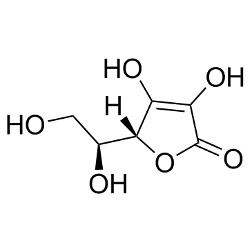 |
Ascorbic acid
CAS:50-81-7 |
|
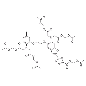 |
Fura-2, AM
CAS:108964-32-5 |
|
 |
Calcium chloride
CAS:10043-52-4 |
|
 |
3-Ethyl-2,4-pentanedione
CAS:1540-34-7 |
|
 |
Osmium tetroxide
CAS:20816-12-0 |
|
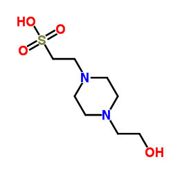 |
HEPES
CAS:7365-45-9 |