Decavanadate binding to a high affinity site near the myosin catalytic centre inhibits F-actin-stimulated myosin ATPase activity.
Teresa Tiago, Manuel Aureliano, Carlos Gutiérrez-Merino
文献索引:Biochemistry 43(18) , 5551-61, (2004)
全文:HTML全文
摘要
Decameric vanadate (V(10)) inhibits the actin-stimulated myosin ATPase activity, noncompetitively with actin or with ATP upon interaction with a high-affinity binding site (K(i) = 0.27 +/- 0.05 microM) in myosin subfragment-1 (S1). The binding of V(10) to S1 can be monitored from titration with V(10) of the fluorescence of S1 labeled at Cys-707 and Cys-697 with N-iodo-acetyl-N'-(5-sulfo-1-naphthyl)ethylenediamine (IAEDANS) or 5-(iodoacetamido) fluorescein, which showed the presence of only one V(10) binding site per monomer with a dissociation constant of 0.16-0.7 microM, indicating that S1 labeling with these dyes produced only a small distortion of the V(10) binding site. The large quenching of AEDANS-labeled S1 fluorescence produced by V(10) indicated that the V(10) binding site is close to Cys-697 and 707. Fluorescence studies demonstrated the following: (i) the binding of V(10) to S1 is not competitive either with actin or with ADP.V(1) or ADP.AlF(4); (ii) the affinity of V(10) for the complex S1/ADP.V(1) and S1/ADP.AlF(4) is 2- and 3-fold lower than for S1; and (iii) it is competitive with the S1 "back door" ligand P(1)P(5)-diadenosine pentaphosphate. A local conformational change in S1 upon binding of V(10) is supported by (i) a decrease of the efficiency of fluorescence energy transfer between eosin-labeled F-actin and fluorescein-labeled S1, and (ii) slower reassociation between S1 and F-actin after ATP hydrolysis. The results are consistent with binding of V(10) to the Walker A motif of ABC ATPases, which in S1 corresponds to conserved regions of the P-loop which form part of the phosphate tube.
相关化合物
| 结构式 | 名称/CAS号 | 分子式 | 全部文献 |
|---|---|---|---|
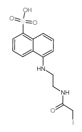 |
N-碘乙酰-N'-(5-磺基-1-萘)乙二胺
CAS:36930-63-9 |
C14H15IN2O4S |
|
Detection of proteins on blot membranes.
2001-05-01 [Curr. Protoc. Protein Sci. Chapter 10 , Unit 10.8, (2001)] |
|
Iron-sulfur cluster biosynthesis: characterization of IscU-I...
2009-08-01 [J. Biol. Inorg. Chem. 14(6) , 829-39, (2009)] |
|
Characterization of the formation of amyloid protofibrils fr...
2006-05-12 [J. Mol. Biol. 358(4) , 935-42, (2006)] |
|
Fluorescence-based peptide labeling and fractionation strate...
2005-07-15 [Anal. Chem. 77(14) , 4495-502, (2005)] |
|
Characterization of low density lipoprotein receptor ligand ...
2006-05-01 [J. Lipid Res. 47(5) , 1091-6, (2006)] |