| 结构式 | 名称/CAS号 | 全部文献 |
|---|---|---|
 |
蔗糖
CAS:57-50-1 |
|
 |
乙醇
CAS:64-17-5 |
|
 |
L-2,4-二氨基丁酸 单盐酸盐
CAS:1482-98-0 |
|
 |
3,3'-二氨基联苯胺
CAS:91-95-2 |
|
 |
锇酸酐
CAS:20816-12-0 |
|
 |
2,2-双-(4-甘胺氧苯)丙烷
CAS:1675-54-3 |
|
 |
苏木精
CAS:517-28-2 |
|
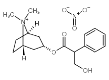 |
甲基硝酸阿托品
CAS:52-88-0 |