Doramapimod (BIRB 796)
Modify Date: 2025-08-20 09:55:18
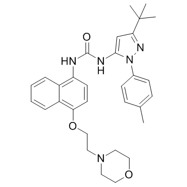
Doramapimod (BIRB 796) structure
|
Common Name | Doramapimod (BIRB 796) | ||
|---|---|---|---|---|
| CAS Number | 285983-48-4 | Molecular Weight | 527.657 | |
| Density | 1.2±0.1 g/cm3 | Boiling Point | 631.6±55.0 °C at 760 mmHg | |
| Molecular Formula | C31H37N5O3 | Melting Point | N/A | |
| MSDS | N/A | Flash Point | 335.8±31.5 °C | |
Use of Doramapimod (BIRB 796)Doramapimod (BIRB 796) is a highly potent p38 MAPK inhibitor with an IC50 of 4 nM. It also inhibits B-Raf with an IC50 of 83 nM. |
| Name | 1-[5-tert-butyl-2-(4-methylphenyl)pyrazol-3-yl]-3-[4-(2-morpholin-4-ylethoxy)naphthalen-1-yl]urea |
|---|---|
| Synonym | More Synonyms |
| Description | Doramapimod (BIRB 796) is a highly potent p38 MAPK inhibitor with an IC50 of 4 nM. It also inhibits B-Raf with an IC50 of 83 nM. |
|---|---|
| Related Catalog | |
| Target |
B-Raf:83.4 nM (IC50) p38α:4 nM (IC50) Abl:14600 nM (IC50) |
| In Vitro | Doramapimod (BIRB 796) is a highly potent inhibitor of p38α, a serine/threonine mitogen activated protein kinase (MAPK) that is usually associated with inflammation because of its role in T-cell proliferation and cytokine production[1]. Doramapimod (BIRB 796) is a picomolar inhibitor of human p38 MAP kinase[2]. HEK293 cells are incubated with different concentrations of Doramapimod (BIRB 796) for 30 min or 2 h prior to stimulation with sorbitol (an osmotic shock) and examined the activation of MAPKAP-K2 by measuring its activity. MAPKAP-K2 activation is inhibited in these cells in a time-dependent manner with an apparent IC50 of 30 nM after 30 min or 8 nM after 2 h of preincubation with Doramapimod[3]. |
| In Vivo | The mean xenograft weigh of Doramapimod (BIRB 796) plus Paclitaxel group (1.14±0.48 g) is lighter than control, Doramapimod or Paclitaxel group (1.84±0.61, 1.80±0.62 and 1.65±0.29 g) (P<0.05) in Athymic nude mice (BALB/c-nu/nu). The inhibition rate of Doramapimod, Paclitaxel and Doramapimod plus Paclitaxel is 1.93%, 9.93% and 38%. Importantly, the combination of Doramapimod and Paclitaxel produces a significant inhibition of tumor growth, compare with control, Paclitaxel or Doramapimod alone[4]. The Doramapimod (BIRB 796) treatment slightly reduces blood pressure (166±7 mm Hg at week 7; P<0.05), whereas SD rats are normotensive (123±3 mm Hg). Despite the reduction in blood pressure, untreated and Doramapimod-treated dTGRs have similar heart weight and cardiac hypertrophy indices (heart-to-tibia ratio), which are significantly higher compare with nontransgenic SD rats (310±6 versus 307±6 versus 206±5 mg/cm, respectively; P<0.05)[5]. |
| Kinase Assay | Binding studies are conducted in a buffer containing 20 mM Bis-Tris Propane, pH 7.0, 2 mM EDTA, 0.01% (w/v) NaN3 and 0.15% (w/v) n-octylglucoside. Kinetic data for the association of SK&F 86002 to p38 MAP kinase is collected on a Kintech fluorescence detector system equipped with a stopped flow controller. The data are fit simultaneously to an appropriate equation describing kinetic binding for a simple one-step binding mechanism. The data for the binding of the fluorescent analog of BIRB 796 is corrected for background fluorescence of unbound ligand. The exchange curve assays are run as two half-reactions using an SLM Aminco Bowman Series 2 Model SQ-340 fluorescence detector. In the first half-reaction, p38 MAP kinase and SK&F 86002 are preincubated for 3 min. In the second half-reaction, p38 MAP kinase is preincubated with Doramapimod for 60 min. A net dissociation of the fluoroprobe is observed for the first half-reaction, and a net association is observed for the second half-reaction. The raw data from both half reactions are fit simultaneously to an equation describing simple competitive inhibition. p38 is preactivated by treatment with constitutively active recombinant MKK6 (prepared by mutagenizing the two activation residues, Ser189 and Thr193, to Glu residues). Activated p38 is purified and used as a source of enzyme in a standard kinase activity assay monitoring the incorporation of radioactive phosphate into recombinant human MAPKAP k2. Cellular assays follow published procedures. Briefly, human THP.1 cells are stimulated with 1 μg/mL LPS, in the presence or absence of compound, followed by the determination of released TNF using a commercial ELISA kitkinase[2]. |
| Cell Assay | Human embryonic kidney (HEK) 293 and HeLa cells are exposed to 0.5 M sorbitol for 30 min or 100 ng/mL EGF for 10 min and then lysed in buffer A (50 mM Tris-HCl, pH 7.5, 1 mM EGTA, 1 mM EDTA, 1 mM sodium orthovanadate, 10 mM sodium fluoride, 50 mM sodium β-glycerophosphate, 5 mM pyrophosphate, 0.27 M sucrose, 0.1 mM phenylmethylsulfonyl fluoride, 1% (v/v) Triton X-100) plus 0.1% (v/v) 2-mercaptoethanol and Complete proteinase inhibitor mixture. Lysates are centrifuged at 18,000× g for 5 min at 4°C, and the supernatants are removed, quick-frozen in liquid nitrogen, and stored at -20°C until use. When required, cells are preincubated for 1 h without or with 10 μM SB 203580 or 10 μM PD 184352 or with different concentrations of Doramapimod for the times indicated in the figures[3]. |
| Animal Admin | Mice[4] Athymic nude mice (BALB/c-nu/nu), 6 to 8 weeks of age and weighing 18 to 24 g, are used. The KBV200-inoculated nude mouse xenograft model is used in this study. In brief, harvested KBV200 cells (2×10⁷ cells/0.2 mL/mouse) are implanted subcutaneously under the shoulder in the nude mice. When the tumors reach a mean diameter of 0.5 cm3, the mice are divided randomly into four groups and are treated with the following regimens: 1) saline solution (every 3 days×5); 2) Paclitaxel (18 mg/kg i.p., every 3 days×5); 3) Doramapimod (10 mg/kg p.o., every 3 days×5); and 4) Paclitaxel (18 mg/kg i.p., every 3 days×5) plus Doramapimod (10 mg/kg p.o., every 3 days×5, administered 1 h before injection of Paclitaxel). The body weights of the animals and the two perpendicular tumor diameters (A and B) are recorded every 3 days, and the tumor volume (V) is estimated according to the following formula: V=(π/6)[(A+B)/2]3. The curves for tumor growth and body weight are drawn according to tumor volume and time of implantation. The mice are anesthetized and killed when the mean tumor weight in the control group is more than 1 g. Tumor tissues are excised from the mice, and their weights are measured. The ratio of growth inhibition (IR) is calculated according to the following formula: IR=[1- (mean tumor weight for experimental group/mean tumor weight for control group)]×100%. Rats[5] Male transgenic dTGRs (RCC Ltd) and age-matched nontransgenic Sprague-Dawley (SD) rats (MDC) are use. 2 different protocols are performed. In protocol 2, untreated dTGR (n=15), dTGR+BIRB796 (30 mg/kg per day in the diet for 3 weeks; n=11), and SD (n=8 each group) rats are analyzed. Systolic blood pressure is measured weekly by tail cuff. Twenty-four-hour urine samples are collected in metabolic cages from weeks 5 to 7. Serum is collected at week 7. Serum creatinine and cystatin C are measured by clinical routine assays. Urinary rat albumin is determined by enzyme-linked immunosorbent assay. The aim of protocol 2 is to focus on electrophysiological alterations and mortality. Untreated dTGR (n=10), dTGR+BIRB796 (n=10), and SD (n=10) rats are studied up to week 8. Cardiac magnetic field mapping (CMFM) is performed at week 7 under isoflurane anesthesia. Echocardiography is performed. |
| References |
| Density | 1.2±0.1 g/cm3 |
|---|---|
| Boiling Point | 631.6±55.0 °C at 760 mmHg |
| Molecular Formula | C31H37N5O3 |
| Molecular Weight | 527.657 |
| Flash Point | 335.8±31.5 °C |
| Exact Mass | 527.289612 |
| PSA | 80.65000 |
| LogP | 6.11 |
| Vapour Pressure | 0.0±1.9 mmHg at 25°C |
| Index of Refraction | 1.620 |
| Storage condition | -20°C |
| 1kv2 |
| BIRB796 |
| 1-[3-tert-Butyl-1-(4-methylphenyl)-1H-pyrazol-5-yl]-3-{4-[2-(morpholin-4-yl)ethoxy]-1-naphthyl}urea |
| S1574_Selleck |
| Kinome_2137 |
| 1-(3-tert-butyl-1-p-tolyl-1H-pyrazol-5-yl)-3-(4-(2-morpholinoethoxy)naphthalen-1-yl)urea |
| L66J BO2- AT6N DOTJ& EMVM- ET5NNJ AR D1& CX1&1&1 |
| doramapimod |
| Urea, N-[3-(1,1-dimethylethyl)-1-(4-methylphenyl)-1H-pyrazol-5-yl]-N'-[4-[2-(4-morpholinyl)ethoxy]-1-naphthalenyl]- |
| 1-[1-(4-Methylphenyl)-3-(2-methyl-2-propanyl)-1H-pyrazol-5-yl]-3-{4-[2-(4-morpholinyl)ethoxy]-1-naphthyl}urea |
| BIRB 796 |
| UNII-HO1A8B3YVV |