| Description |
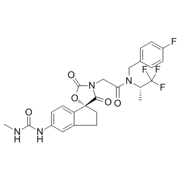
A-485 is a potent and selective catalytic inhibitor of p300/CBP with IC50s of 9.8 nM and 2.6 nM for p300 and CBP, respectively.
|
| Related Catalog |
|
| Target |
IC50: 9.8 nM (p300), 2.6 nM (CBP)[1]
|
| In Vitro |
A three-hour treatment of prostate adenocarcinoma PC-3 cells with A-485 results in a dose-dependent decrease in H3K27Ac, with a half maximal effective concentration (EC50) of 73 nM. Treatment with A-485 does not alter p300 or CBP protein levels. The broadest sensitivity is observed in haematological tumours, where A-485 exhibits potent activity in most multiple myeloma cell lines, and in a subset of acute myeloid leukaemia lines and non-Hodgkin’s lymphoma lines. A-485 induces a comparable decrease in H3K27Ac in all five prostate cancer cell lines. Treatment of the androgen-dependent LnCaP-FGC cell line with A-485 attenuates dihydrotestosterone (DHT) stimulated prostate-specific antigen (PSA) expression[1].
|
| In Vivo |
After tumours are established in SCID male mice, twice daily intraperitoneal injections of A-485 induce 54% tumour growth inhibition after 21 days of dosing (P<0.005 as compare to vehicle control). In addition, in tumour-bearing animals, dosing with A-485 for seven days induces a decrease in the mRNA levels of MYC and the AR-dependent gene SLC45A3 at three hours post-dosing, and (for MYC) a decrease in the protein level, indicating that A-485 inhibits p300-mediated transcriptional activity in vivo. However, at 16 hours post-dosing on the seventh day, A-485 drug levels in the plasma and tumour are decreased as compare to 3 hours. A-485 induces a moderate 9% body weight loss, and the animals recover rapidly upon completion of the A-485 dosing regimen[1].
|
| Cell Assay |
Cell lines are plated in 96 well or 384 well plates and allowed to adhere for 24 h. The cells are then treated with A-485 for 3, 4, or 5 days. Experiments are run in triplicate and the fraction of viable cells is determined using the Cell Viability Assay according to the manufacturer’s recommendations. For Thymidine incorporation assays, cells are treated with A-485 for 1, 2, 3, or 4 days. Twenty four hours prior to the time point, tritiated thymidine is added and cells are incubated for an additional 24 h. Genomic DNA is then isolated on filter plates[1].
|
| Animal Admin |
The LuCap-77 CR prostate PDX model is used in this study. Donor tumors are dissociated and injected as a brie (1:2) into the right flank of 16 week old male C.B.-17 SCID mice on day 0 in a volume of 0.2 mL. Tumors are size matched on day 26 post-inoculation with a mean tumor volume of 211±3 (SEM) mm3 with dosing beginning on day 28. Mice are randomized into treatment groups using Studylog software based on tumor volume. LuCap-77 CR xenograft tumors are established in SCID mice and animals are dosed with A-485 as for 7 days. Three hours post the final dose, tumors are harvested and snap frozen on dry ice[1].
|
| References |
[1]. Lasko LM, et al. Discovery of a selective catalytic p300/CBP inhibitor that targets lineage-specific tumours. Nature. 2017 Oct 5;550(7674):128-132.
|
