| Description |
Erlotinib is a medication for the treatment of non-small cell lung cancer. It inhibits purified EGFR kinase with an IC50 of 2 nM.
|
| Related Catalog |
|
| Target |
EGFR:2 nM (IC50, Cell Free Assay)
|
| In Vitro |
Erlotinib (CP-358,774) is also a potent inhibitor of the recombinant intracellular (kinase) domain of the EGFR, with an IC50 of 1 nM. The proliferation of DiFi cells is strongly inhibited by Erlotinib with an IC50 of 100 nM for an 8-day proliferation assay[1]. The combination of B-DIM and Erlotinib (2 μM) results in a significant inhibition of colony formation in BxPC-3 cells when compared with either agent alone. The combination of B-DIM and Erlotinib (2 μM) results in a significant induction of apoptosis only in BxPC-3 cells when compare with the apoptotic effect of either agent alone[2].
|
| In Vivo |
Under the experimental conditions, the combination of B-DIM and Erlotinib (50 mg/kg, i.p.) treatment shows significant decrease (P <0.01) in tumor weight compared with untreated control[2]. Erlotinib (20 mg/kg, p.o.) significantly attenuates Cisplatin (CP)-induced body weight (BW) loss when compared to the CP+vehicle (V) rats (P<0.05). Erlotinib treatment significantly improves renal function in CP-N(normal control group, NC) rats. The CP+Erlotinib (E) rats show significant reduction of the levels of Serum creatinine (s-Cr) (P<0.05), blood urea nitrogen (BUN) (P<0.05), urinary N-acetyl-β-D-glucosaminidase (NAG) index (P<0.05), and significant increase of urine volume (UV) (P<0.05) and Cr clearance (Ccr) (P<0.05) compare to the CP+V rats[3]
|
| Kinase Assay |
The kinase reaction is performed in 50 μL of 50 mM HEPES (pH 7.3), containing 125 mM NaCl, 24 mM MgCl2, 0.1 mM Na3VO4, 20 μM ATP, 1.6 μg/mL EGF, and 15 ng of EGFR, affinity purified from A431 cell membranes. The compound in DMSO is added to give a final DMSO concentration of 2.5%. Phosphorylation is initiated by addition of ATP and proceeded for 8 mm at room temperature, with constant shaking. The kinase reaction is terminated by aspiration of the reaction mixture and is washed 4 times with wash buffer. Phosphorylated PGT is measured by 25 mim of incubation with 50 μL per well HRP-conjugated PY54 antiphosphotyrosine antibody, diluted to 0.2 μg/mL in blocking buffer (3% BSA and 0.05% Tween 20 in PBS). Antibody is removed by aspiration, and the plate is washed 4 times with wash buffer. The colonmetric signal is developed by addition of TMB Microwell Peroxidase Substrate, 50 μL per well, and stopped by the addition of 0.09 M sulfuric acid, 50μL per well. Phosphotyrosine is estimated by measurement of absorbance at 450 nm. The signal for controls is typically 0.6-1.2 absorbance units, with essentially no back ground in wells without AlP, EGFR, or POT and is proportional to the time of incubation for 10 mm[1].
|
| Cell Assay |
To test the viability of cells treated with B-DIM, Erlotinib, or the combination, BxPC-3 and MIAPaCa cells are plated (3,000-5,000 per well) in a 96-well plate and incubated overnight at 37°C. A range of concentrations for both B-DIM (10-50 µM) and Erlotinib (1-5 µM) is initially tested. Based on the initial results, the concentration of B-DIM (20 µM) and Erlotinib (2 µM) are chosen for all assays. The effects of B-DIM (20 µM), Erlotinib (2 µM), and the combination on BxPC-3 and MIAPaCa cells are determined by the standard MTT assay after 72 h and is repeated three times. The color intensity is measured by a Tecan microplate fluorometer at 595 nm. DMSO-treated cells are considered to be the untreated control and assigned a value of 100%. In addition to the above assay, we have also done clonogenic assay for assessing the effects of treatment[2].
|
| Animal Admin |
Mice[2] Female ICR-SCID (6-7 weeks old) mice are randomized into the following treatment groups (n=7): (a) untreated control; (b) only B-DIM (50 mg/kg body weight), intragastric once every day; (c) Erlotinib (50 mg/kg body weight), everyday i.p. for 15 days; and (d) B-DIM and Erlotinib, following schedule as for individual treatments. All mice are killed on day 3 following last dose of treatment, and their body weight is determined. One part of the tissue is rapidly frozen in liquid nitrogen and stored at −70°C for future use and the other part is fixed in formalin and processed for paraffin block. H&E staining of fixed tissue section is used to confirm the presence of tumor(s) in each pancreas. Rats[3] Six-week-old male Sprague-Dawley (SD) rats weighing 180 to 210 g are used. Cisplatin (CP) is freshly prepared in saline at a concentration of 1 mg/mL and then injected intraperitoneally in SD rats (n=28) at a dose of 7 mg/kg on day 0. To investigate the effect of Erlotinib, 28 CP-N rats are divided into two groups. Separate groups (n=14) each of animals are administered with either Erlotinib (20 mg/kg) (CP+E, n=14) or vehicle (CP+V, n=14) daily by oral gavage from day -1 (24 hours prior to the CP injection) to day 3. Vehicle-treated groups receive an equivalent volume of saline. Five male SD rats at the age of 6 weeks are used as a normal control group (NC, n=5). The NC rats are given an equivalent volume of saline daily by oral gavage from day -1 to day 3. At day 4 (96 hours after CP injection), each rat is anesthetized and sacrificed by exsanguination after the cardiac puncture; blood is collected by cardiac puncture and kidneys are collected. Renal tissue is divided; separate portions are snap-frozen in liquid nitrogen or fixed in 2% paraformaldehyde/phosphate-buffered saline (PBS) for later use. All surgery is performed under diethyl ether gas anesthesia, and all efforts are made to minimize suffering.
|
| References |
[1]. Moyer JD, et al. Induction of apoptosis and cell cycle arrest by CP-358,774, an inhibitor of epidermal growth factor receptor tyrosine kinase. Cancer Res. 1997, 57(21), 4838-4848. [2]. Ali S, et al. Apoptosis-inducing effect of erlotinib is potentiated by 3,3'-diindolylmethane in vitro and in vivo using an orthotopic model of pancreatic cancer. Mol Cancer Ther, 2008, 7(6), 1708-1719. [3]. Wada Y, et al. Epidermal growth factor receptor inhibition with erlotinib partially prevents cisplatin-induced nephrotoxicity in rats. PLoS One. 2014 Nov 12;9(11):e111728.
|

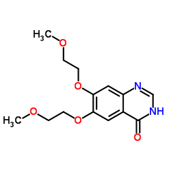 CAS#:179688-29-0
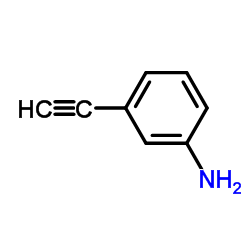 CAS#:54060-30-9
![N'-[2-cyano-4,5-bis(2-methoxyethoxy)phenyl]-N,N-dimethylmethanimidamide Structure](https://image.chemsrc.com/caspic/215/950596-59-5.png) CAS#:950596-59-5
CAS#:950596-59-5 CAS#:183319-69-9
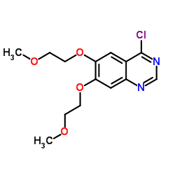 CAS#:183322-18-1
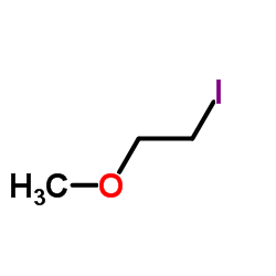 CAS#:4296-15-5
 CAS#:950596-58-4