| In Vitro |
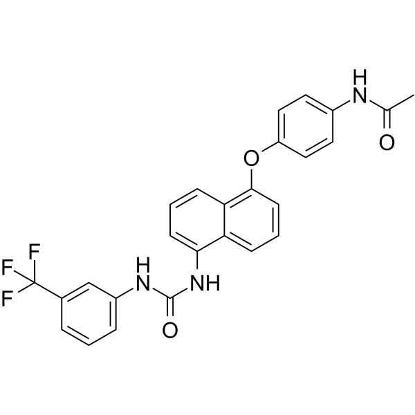
VS 8 (Compound VS 8) (0.01-100 µM, 24 h) shows potent anti-proliferative activity against MCF-7, MDA-MB-231, Hep G2, and HUVECs cells[1]. VS 8 induces early apoptosis in MDA-MB-231 (1413 nM, 72 h), Hep G2 (257.80 nM, 24 h), and HUVECs (1954 nM, 24 h) cells[1]. VS 8 is shown to be a pro-oxidant molecule that enhances the ROS level in Hep G2 cells[1]. VS 8 inhibits wound healing and migration of MCF-7 cancer cells[1]. VS 8 downregulates human vascular endothelial growth factor (hVEGF) and hVEGFR-2 expression in HUVECs[1]. VS 8 (257.80 nM, 48 h) arrests cell cycle at ‘G0/G1’ and ‘S’ phase in CD44+ and CD133+ CSCs, respectively[1]. VS 8 inhibits TGF-β-induced epithelial-mesenchymal transition (EMT) in hepatocellular carcinoma by the upregulation of E-cadherin and the suppression of vimentin and SNAIL[1]. Cell Proliferation Assay[1] Cell Line: MCF-7, MDA-MB-231, Hep G2, and HUVECs cells Concentration: 0.01, 0.1, 1, 10, 50, and 100 µM Incubation Time: 24 h Result: Showed significantly potent anti-proliferative activity against all the selected cell lines in a dose-dependent manner, with IC50 values of 953.30, 1413, 257.80, and 1954 nM against MCF-7, MDA-MB-231, Hep G2, and HUVECs cells. Apoptosis Analysis[1] Cell Line: MDA-MB-231, Hep G2, and HUVECs cells Concentration: 1413, 257.80, and 1954 nM for MDA-MB-231, Hep G2, and HUVECs cells, respectively. Incubation Time: 72 h for MDA-MB-231 cells; 24 h for Hep G2 and HUVECs cells Result: Resulted in high population of early apoptotic MDA-MB-231 cells (68.34 ± 0.18%). A significant increase in % apoptotic index (~86.66%) was observed in Hep G2 cells. The percentage of early apoptotic cells were found to be ~37.53% in HUVECs cells. Cell Cycle Analysis[1] Cell Line: CD44+ and CD133+ CSCs isolated from Hep G2 cells Concentration: 257.80 nM Incubation Time: 48 h Result: Arrested cell cycle at ‘G0/G1’ and ‘S’ phase in CD44+ and CD133+ CSCs, respectively.
|
| In Vivo |
VS 8 inhibits angiogenesis in the chick chorioallantoic membrane without oral toxicity[1]. Animal Model: Male Wistar rats (180-220 gm)[1] Dosage: 5 mg/kg Administration: Oral administration (Pharmacokinetic Analysis) Result: Pharmacokinetic parameters for VS 8 in rats after administration of oral dose (5 mg/ kg) [1] Pharmacokinetic parameters Unit Value Cmax μg/mL 39.7193 ± 0.36 Tmax hrs 6 ± 0 AUC(0-72) mg/mL*hrs 621.3236 ± 1.843 AUC(0-∞) mg/mL*hrs 625.2219 ± 1.864 AUMC(0-∞) (mg/mL*hrs2) 8929.284 ± 72.85 MRT hrs 14.2817 ± 0.102 t1/2 hrs 11.9277 ± 0.324 Data represented as mean ± SD (n = 3); t1/2, Half-Life; Cmax, Maximum Observed Concentration; Tmax, Maximum Observed Time; AUC, Area Under Curve; AUMC Area Under Movement Curve, MRT, Mean Residence Time.
|
