| Description |
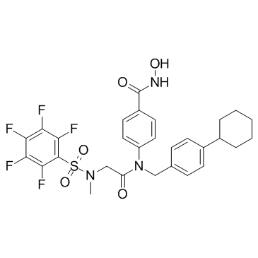
SH5-07 is a hydroxamic acid based Stat3 inhibitor with an IC50 of 3.9 μM in in vitro assay.
|
| Related Catalog |
|
| Target |
STAT3:3.9 μM (IC50)
|
| In Vitro |
SH5-07 is a hydroxamic acid analog of BP-1-102. SH5-07 dose-dependently inhibits Stat3 activity with an IC50 of 3.9±0.6 μM in in vitro assay. It preferentially inhibits Stat3:Stat3 DNA-binding activity, ahead of inhibiting Stat1:Stat3 activity, with minimal effects on Stat1:Stat1 activity. SH5-07 binds Stat3, disrupts Stat3 association with growth factor receptor, and thereby inhibits Stat3 phosphorylation. It induces antitumor cell effects against malignant cells harboring constitutively-active Stat3. SH5-07 inhibits the expression of known Stat3-regulated genes. Bcl-2, Bcl-xL, c-Myc, Survivin, Cyclin D1 and Mcl-1 expression is reduced in response to 24 h, 5 μM SH5-07 treatment[1].
|
| In Vivo |
Tail vein injection or oral gavage delivery of SH5-07 or SH4-54 inhibits growth of 90-150 mm3 established subcutaneous mouse xenografts of human glioma (U251MG) and breast (MDA-MB-231) tumors that harbor aberrantly-active Stat3, associated with decreased c-Myc, Mcl-1 and Cyclin D1 expression. No significant changes in body weights, blood cell counts, or the gross anatomy of organs, or obvious signs of toxicity are observed[1].
|
| Cell Assay |
Cells are treated with 0-8 μM agent for 24-48 h. For cell cycle profile analysis, cells are harvested and fixed with 70% ice-cold ethanol and stained with propidium iodide (PI). For apoptosis analysis, cells are collected and stained with FITC-Annexin V using Apoptosis Detection Kit. Both the DNA content of cells and the Annexin V-positive cells are analyzed by FACScan flow cytometer. Cell cycle phase distribution is analyzed using the Cell-Fit program. Data acquisition is gated to exclude cell doublets[1].
|
| Animal Admin |
Mice: Mice are injected subcutaneously in the left flank area with U251MG cells in 200 μL of PBS/Matrigel matrix, or MDA-MB-231 cells in 100 μL of PBS. Mice with tumors of 90-150 mm3 (MDA-MB-231) or 150 mm3 (U251MG) are grouped for identical mean tumor sizes, administered 3, 5 or 6 mg/kg SH5-07 or SH4-54 via oral gavage daily or tail vein injection every 2 or 3 days, and monitored every 3-7 days. Tumor sizes are measured with calipers and converted to tumor volume[1].
|
| References |
[1]. Yue P,et al. Hydroxamic Acid and Benzoic Acid-Based STAT3 Inhibitors Suppress Human Glioma and Breast Cancer Phenotypes In Vitro and In Vivo. Cancer Res. 2016 Feb 1;76(3):652-63.
|