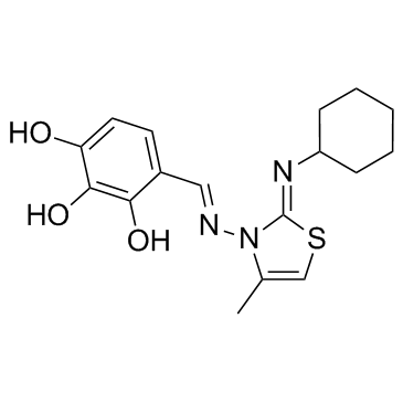
| Cell Assay |
The IC50 value, defined as the concentration that reduces the global growth of cells by 50%, is determined for the apoptosis inhibitors ABT-737 and MIM-1, individually, for the human astrocyte (HA) and human GB (T98G) cell lines. The apoptosis inhibitor concentrations and treatment time periods are selected experimentally according to preliminary experiments. The final ABT-737 treatment is performed with 10-fold increasing concentrations in the range of 0.001-100 µM, and the final MIM-1 treatment is performed with 4-fold increasing concentrations in the range of 0.4-400 µM, for 48 h. The biochemical colorimetric MTT assay, based on the enzymatic conversion of MTT to a violet formazan salt, is used to assess the viability of the HA and T98G cells. Briefly, the cells in culture medium are seeded (3.5×10³ cells/well for the HA cell line, 3.0×10³ cells/well for T98G cell line) in 96-well microtiter plates. On the third day, the medium is changed to culture medium supplemented with the apoptosis inhibitor ABT-737 or MIM-1 at varied concentrations and incubation continued for another two days. After the treatment with the apoptosis inhibitors, cells are rinsed once with Dulbeccos phosphate buffer saline (DPBS) and further incubated in medium supplemented with 0.5 mg/mL MTT in a humidified atmosphere for 6 h. During a subsequent incubation for 16 h in medium containing SDS [5% (w/v)], the precipitated formazan, the amount of which is proportional to the number of live cells, is solubilized. The absorbance of the formazan-containing solution is measured at 540 nm using an ELISA plate reader. The absorbance is also determined for the medium of the control cells not exposed to the apoptosis inhibitors. The percentage of cell viability is calculated relative to the untreated control cells. The IC50 values are determined for both human brain cell lines after individual apoptosis inhibitor treatment.
|
