| Description |
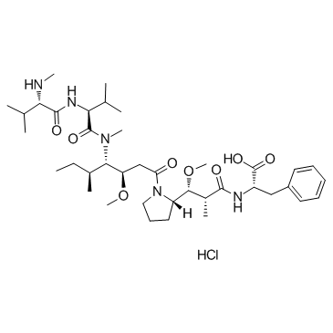
MMAF hydrochloride is an antitubulin agent that inhibit cell division; inhibits H3397 cell growth with an IC50 of 105 nM.
|
| Related Catalog |
|
| Target |
IC50: 119 nM (Cytotoxicity, Karpas 299 cell), 105 nM (Cytotoxicity, H3396 cell), 257 nM (Cytotoxicity, 786-O cell), 200 nM (Cytotoxicity, Caki-1, cell)[1]
|
| In Vitro |
MMAF shows in vitro cytotoxicity against a panel of cell lines. The IC50 values for Karpas 299, H3396, 786-O and Caki-1 are 119, 105, 257, and 200 nM, respectively. Targeted MMAF is much more potent than the free drug, and that cAC10 conjugates of MMAF display pronounced activities. On a molar basis, the cAC10-L1-MMAF4 is an average of over 2200-fold more potent than free MMAF and is active on all the CD30-positive cell lines tested[1].
|
| In Vivo |
The maximum tolerated dose in mice of MMAF (>16 mg/kg) is much higher than MMAE (1 mg/kg). cAC10-L1-MMAF4 has an MTD of 50 mg/kg in mice and 15 mg/kg in rats. The corresponding cAC10-L4-MMAF4 ADC was much less toxic, having MTDs in mice and rats of >150 mg/ kg and 90 mg/kg in rats, respectively[1].
|
| Cell Assay |
Cells are treated with serial dilutions of test molecules and incubated 4-6 days depending on cell line. Assessment of cellular growth and data reduction to generate IC50 values is done using Alamar Blue dye reduction assay[1].
|
| Animal Admin |
Mice: When subcutaneous Karpas 299 tumor size reaches 300 mm3, three animals per group receives one injection of 10 mg antibody component/kg body weight of either cAC10-L1-MMAF4 or cBR96-L1-MMAF4 intravenously. Tumors are then removed and placed in optimal cutting temperature compound, and 5 μm-thin frozen tissue sections are stained using immunohistochemistry evaluation[1].
|
| References |
[1]. Doronina SO, et al. Enhanced activity of monomethylauristatin F through monoclonal antibody delivery: effects of linker technology on efficacy and toxicity. Bioconjug Chem. 2006 Jan-Feb;17(1):114-24.
|