178119-00-1
178119-00-1 structure
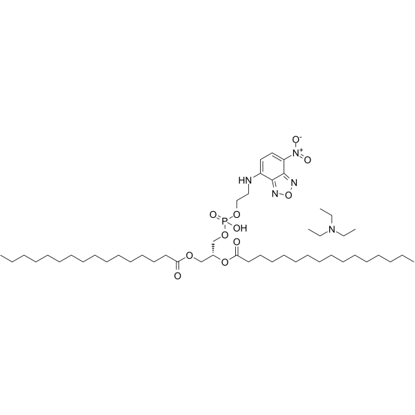
- Name: NBD-PE [N-(7-Nitrobenz-2-oxa-1,3-diazol-4-yl)-1,2-dihexadecanoyl-sn-glycero-3-phosphoethanolamine, triethylammonium salt]
- Chemical Name: nbd-pe
- CAS Number: 178119-00-1
- Molecular Formula: C49H90N5O11P
- Molecular Weight: 956.24000
- Catalog: Research Areas Others
- Create Date: 2016-08-21 16:38:37
- Modify Date: 2025-08-25 23:33:34
-
NBD-PE is an effective lipid fluorescent probe (Excitation/Emission: 465/535 nm; Color: Green). NBD-PE offers a wide array of applications in membrane and cell biology[1][2][3].
| Name | nbd-pe |
|---|
| Description | NBD-PE is an effective lipid fluorescent probe (Excitation/Emission: 465/535 nm; Color: Green). NBD-PE offers a wide array of applications in membrane and cell biology[1][2][3]. |
|---|---|
| Related Catalog | |
| In Vitro | Guidelines (Following is our recommended protocol. This protocol only provides a guideline, and should be modified according to your specific needs). Assay of phospholipid transfer activity[3]: 1. The reaction mixture containes 2 mg mitochondrial protein, 0.02 μmol of NBD-PE, 0.5 mmol of Tris-HCl, pH 7.4, and 0-200 μg of pH 5.l-supernatant protein. 2. Samples are incubated at 37°C for 1 hour with gentle agitation. 3. The reaction is terminated by placing samples in an ice bath for 10 min, followed by centrifugation at 15,000 g for 5 min. 4. The supernatants are decanted, allowed to come to room temperature, and the absorbance (460 nm) and the relative fluorescence at 535 nm (excitation at 465 nm) are determined. 5. Blanks containing only NBD-PE and exchange protein are run concomitantly to correct for possible interaction between NBD-PE and exchange protein. 6. The percent exchange of NBD-PE is calculated from the differences between the absorbances or relative fluorescence in the supernatant fractions in the presence and absence of exchange protein. |
| References |
| Molecular Formula | C49H90N5O11P |
|---|---|
| Molecular Weight | 956.24000 |
| Exact Mass | 955.63700 |
| PSA | 218.18000 |
| LogP | 14.03840 |
| Storage condition | -20°C |