A stopped-flow kinetic study of the interaction of potential-sensitive oxonol dyes with lipid vesicles.
R J Clarke, H J Apell
Index: Biophys. Chem. 34(3) , 225-37, (1989)
Full Text: HTML
Abstract
The interaction of the dyes oxonol V and oxonol VI with unilamellar dioleoylphosphatidylcholine vesicles was investigated using a fluorescence stopped-flow technique. On mixing with the vesicles, both dyes exhibit an increase in their fluorescence, which occurs in two phases. According to the dependence of the reciprocal relaxation time on vesicle concentration, the rapid phase appears to be due to a second-order binding of the dye to the lipid membrane, which is very close to being diffusion-controlled. The slow phase is almost independent of vesicle concentration, and it is suggested that this may be due to a change in dye conformation or position within the membrane, possibly diffusion across the membrane to the internal monolayer. The response times of the dyes to a rapid jump in the membrane potential has also been investigated. Oxonol VI was found to respond to the potential change in less than 1 s, whereas oxonol required several minutes. This has been attributed to lower mobility of oxonol V within the lipid membrane.
Related Compounds
| Structure | Name/CAS No. | Molecular Formula | Articles |
|---|---|---|---|
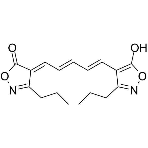 |
Oxonol VI
CAS:64724-75-0 |
C17H20N2O4 |
|
Conserved amino acid residues of the NuoD segment important ...
2015-01-27 [Biochemistry 54(3) , 753-64, (2015)] |
|
ATP-dependent spectral response of oxonol VI in an ATP-Pi ex...
1984-08-31 [Biochim. Biophys. Acta 766(2) , 375-85, (1984)] |
|
Effects of Cd2+ on ATP-driven membrane potential in beef hea...
1984-01-01 [Membr. Biochem. 5(3) , 225-41, (1984)] |
|
Na+-pyrophosphatase: a novel primary sodium pump.
2007-07-31 [Biochemistry 46(30) , 8872-8, (2007)] |
|
Electrophysiological study with oxonol VI of passive NO3- tr...
1999-01-01 [Biophys. J. 76(1 Pt 1) , 360-73, (1999)] |