Microtubule-dependent motility and orientation of the cortical endoplasmic reticulum in elongating characean internodal cells.
Ilse Foissner, Diedrik Menzel, Geoffrey O Wasteneys
Index: Cell Motil. Cytoskeleton 66(3) , 142-55, (2009)
Full Text: HTML
Abstract
Motility of the endoplasmic reticulum (ER) is predominantly microtubule- dependent in animal cells but thought to be entirely actomyosin-dependent in plant cells. Using live cell imaging and transmission electron microscopy to examine ER motility and structural organization in giant internodal cells of characean algae, we discovered that at the onset of cell elongation, the cortical ER situated near the plasma membrane formed a tight meshwork of predominantly transverse ER tubules that frequently coaligned with microtubules. Microtubule depolymerization increased mesh size and decreased the dynamics of the cortical ER. In contrast, perturbing the cortical actin array with cytochalasins did not affect the transverse orientation but decreased mesh size and increased ER dynamics. Our data suggest that myosin-dependent ER motility is confined to the ER strands in the streaming endoplasm, while the more sedate cortical ER uses microtubule-based mechanisms for organization and motility during early stages of cell elongation. We show further that the ER has an inherent, NEM-sensitive dynamics which can be altered via interaction with the cytoskeleton and that tubule formation and fusion events are cytoskeleton-independent.2009 Wiley-Liss, Inc.
Related Compounds
| Structure | Name/CAS No. | Molecular Formula | Articles |
|---|---|---|---|
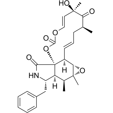 |
Cytochalasin E
CAS:36011-19-5 |
C28H33NO7 |
|
Excessive expression of the plant kinesin TBK5 converts cort...
2007-05-01 [Plant Cell Physiol. 48(5) , 753-61, (2007)] |
|
Cytochalasin E alters the cytoskeleton and decreases ENaC ac...
2014-07-01 [Am. J. Physiol. Renal Physiol. 307(1) , F86-95, (2014)] |
|
Biosynthesis: A carbonate architect emerges.
2014-07-01 [Nat. Chem. Biol. 10(7) , 486-7, (2014)] |
|
A carbonate-forming Baeyer-Villiger monooxygenase.
2014-07-01 [Nat. Chem. Biol. 10(7) , 552-4, (2014)] |
|
Effects of medium calcium, and agents affecting cytoskeletal...
2012-06-01 [J. Cell. Biochem. 113(6) , 1915-25, (2012)] |