Molecular imaging of retinal gliosis in transgenic mice induced by kainic acid neurotoxicity.
Gideon Ho, Saravana Kumar, Xiao Shan Min, Yin Ling Kng, Ming Yang Loh, Shujun Gao, Lang Zhuo
Index: Invest. Ophthalmol. Vis. Sci. 50 , 2459-64, (2009)
Full Text: HTML
Abstract
Gliosis is a universal response of the central nervous system to diverse insults. Here the authors aimed to develop a noninvasive fluorescence system to monitor and quantify retinal gliosis in real time.Transgenic mice expressing green fluorescent protein (GFP) under the control of the glial fibrillary acidic protein promoter were treated with excitatory neurotoxicant kainic acid (KA) through a single intraperitoneal injection to induce gliosis in the brain and the retina. The expression of the GFAP-GFP transgene as a surrogate reporter for gliosis was noninvasively and longitudinally imaged with a confocal scanning laser ophthalmoscope for 2 weeks to monitor the progression of gliosis.The authors demonstrated that KA-induced gliosis (an elevation in GFP fluorescence intensity [FI]) could be noninvasively detected starting on day 3 and that it peaked on day 7, as quantified for the optic disc astrocytes. A significant increase in the FI in retinal glial cells was also visible on the processed images. Immunohistochemistry in defined regions of the brain (hippocampal CA1, CA3, dentate gyrus) known to be affected by KA neurotoxicity showed that severe gliosis in these regions occurred at day 7, when retinal gliosis peaked.The current real-time fluorescent imaging method described here is a powerful preclinical tool to directly monitor retinal gliosis caused by various retinopathies. In addition, this molecular imaging method should be useful in assessing retinal neurotoxicity and in therapeutic development in a preclinical setting.
Related Compounds
| Structure | Name/CAS No. | Molecular Formula | Articles |
|---|---|---|---|
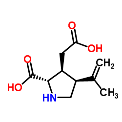 |
Kainic acid hydrate
CAS:58002-62-3 |
C10H15NO4 |
|
MicroRNA-124 and -137 cooperativity controls caspase-3 activ...
2015-01-01 [Sci. Rep. 5 , 12448, (2015)] |
|
Nodule excitability in an animal model of periventricular no...
2015-04-01 [Epilepsia 56(4) , 626-35, (2015)] |
|
Plasticity of calcium signaling cascades in human embryonic ...
2013-05-15 [Stem Cells Dev. 22(10) , 1506-21, (2013)] |
|
CNS oxidative stress associated with the kainic acid rodent ...
2000-03-01 [Epilepsy Res. 39 , 63-71, (2000)] |
|
Neuroprotection requires the functions of the RNA-binding pr...
2015-05-01 [Cell Death Differ. 22(5) , 703-18, (2015)] |