| Structure | Name/CAS No. | Articles |
|---|---|---|
 |
Magnesium choride
CAS:7786-30-3 |
|
 |
L-Glutamic acid potassium salt monohydrate
CAS:6382-01-0 |
|
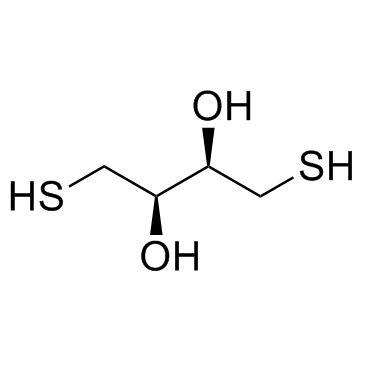 |
DL-Dithiothreitol
CAS:3483-12-3 |