| Structure | Name/CAS No. | Articles |
|---|---|---|
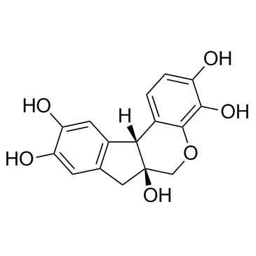 |
Hematoxylin
CAS:517-28-2 |
|
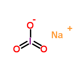 |
Sodium iodate
CAS:7681-55-2 |
|
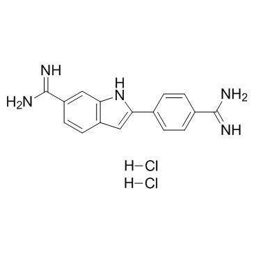 |
4',6-Diamidino-2-phenylindole dihydrochloride
CAS:28718-90-3 |