| Structure | Name/CAS No. | Articles |
|---|---|---|
 |
chromium potassium sulfate
CAS:10141-00-1 |
|
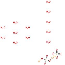 |
Chromium potassium sulfate dodecahydrate
CAS:7788-99-0 |