| Structure | Name/CAS No. | Articles |
|---|---|---|
 |
Arachidic acid
CAS:506-30-9 |
|
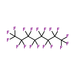 |
Perfluorooctane
CAS:307-34-6 |