| Structure | Name/CAS No. | Articles |
|---|---|---|
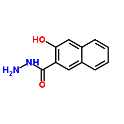 |
3-Hydroxy-2-naphthohydrazide
CAS:5341-58-2 |
|
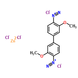 |
Fast Blue B Salt
CAS:14263-94-6 |