Role of 20-HETE in the hypoxia-induced activation of Ca2+-activated K+ channel currents in rat cerebral arterial muscle cells.
Debebe Gebremedhin, Ken Yamaura, David R Harder
Index: Am. J. Physiol. Heart Circ. Physiol. 294(1) , H107-20, (2008)
Full Text: HTML
Abstract
The mechanism of sensing hypoxia and hypoxia-induced activation of cerebral arterial Ca(2+)-activated K(+) (K(Ca)) channel currents and vasodilation is not known. We investigated the roles of the cytochrome P-450 4A (CYP 4A) omega-hydroxylase metabolite of arachidonic acid, 20-hydroxyeicosatetraenoic acid (20-HETE), and generation of superoxide in the hypoxia-evoked activation of the K(Ca) channel current in rat cerebral arterial muscle cells (CAMCs) and cerebral vasodilation. Patch-clamp analysis of K(+) channel current identified a voltage- and Ca(2+)-dependent 238 +/- 21-pS unitary K(+) currents that are inhibitable by tetraethylammonium (TEA, 1 mM) or iberiotoxin (100 nM). Hypoxia (<2% O(2)) reversibly enhanced the open-state probability (NP(o)) of the 238-pS unitary K(Ca) current in cell-attached patches. This effect of hypoxia was not observed on unitary K(Ca) currents recorded from either excised inside-out or outside-out membrane patches. Inhibition of CYP 4A omega-hydroxylase activity increased the NP(o) of K(Ca) single-channel current. Hypoxia reduced the basal endogenous level of 20-HETE by 47 +/- 3% as well as catalytic formation of 20-HETE in cerebral arterial muscle homogenates as determined by liquid chromatography-mass spectrometry analysis. The concentration of authentic 20-HETE was reduced when incubated with the superoxide donor KO(2). Exogenous 20-HETE (100 nM) attenuated the hypoxia-induced activation of the K(Ca) current in CAMCs. Hypoxia did not augment the increase in NP(o) of K(Ca) channel current induced by suicide inhibition of endogenous CYP 4A omega-hydroxylase activity with 17-octadecynoic acid. In pressure (80 mmHg)-constricted cerebral arterial segments, hypoxia induced dilation that was partly attenuated by 20-HETE or by the K(Ca) channel blocker TEA. Exposure to hypoxia caused the generation of intracellular superoxide as evidenced by intense staining of arterial muscle with the fluorescent probe hydroethidine, by quantitation using fluorescent HPLC analysis, and by attenuation of the hypoxia-induced activation of the K(Ca) channel current by superoxide dismutation. These results suggest that the exposure of CAMCs to hypoxia results in the generation of superoxide and reduction in endogenous level of 20-HETE that may account for the hypoxia-induced activation of arterial K(Ca) channel currents and cerebral vasodilation.
Related Compounds
| Structure | Name/CAS No. | Molecular Formula | Articles |
|---|---|---|---|
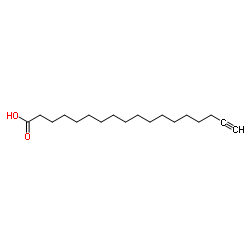 |
17-ODYA
CAS:34450-18-5 |
C18H32O2 | |
 |
Potassium superoxide
CAS:12030-88-5 |
KO2 |
|
Bioorthogonal mimetics of palmitoyl-CoA and myristoyl-CoA an...
2015-06-01 [Proteomics 15 , 2066-77, (2015)] |
|
Interaction of epoxyeicosatrienoic acids and adipocyte fatty...
2015-05-01 [Int. J. Obes. 39 , 755-61, (2015)] |
|
Species differences in intestinal metabolic activities of cy...
2011-06-01 [Drug Metab. Pharmacokinet. 26(3) , 300-6, (2011)] |
|
Arachidonic acid-dependent gene regulation during preadipocy...
2014-12-01 [J. Lipid Res. 55(12) , 2479-90, (2014)] |
|
Changes in renal haemodynamics induced by indomethacin in th...
2004-10-01 [Clin. Exp. Pharmacol. Physiol. 31(10) , 683-90, (2004)] |