| Structure | Name/CAS No. | Articles |
|---|---|---|
 |
Zinc
CAS:7440-66-6 |
|
 |
L-cysteine
CAS:52-90-4 |
|
 |
L-glutamic acid
CAS:56-86-0 |
|
 |
D(-)-Glutamic acid
CAS:6893-26-1 |
|
 |
L-Glutamic acid:Hcl (17O4)
CAS:138-15-8 |
|
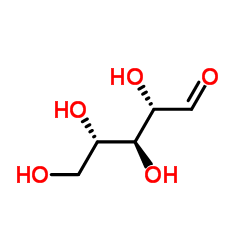 |
L-(+)-Ribose
CAS:147-81-9 |