| Structure | Name/CAS No. | Articles |
|---|---|---|
 |
Sodium Fluoride
CAS:7681-49-4 |
|
 |
Trometamol
CAS:77-86-1 |
|
 |
Sorbitol
CAS:50-70-4 |
|
 |
Sodium orthovanadate
CAS:13721-39-6 |
|
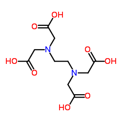 |
Ethylenediaminetetraacetic acid
CAS:60-00-4 |
|
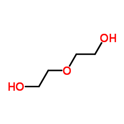 |
Diethylene glycol
CAS:111-46-6 |