Multiparameter MRI assessment of normal-appearing and diseased vertebral bone marrow.
Andreas Biffar, Andrea Baur-Melnyk, Gerwin P Schmidt, Maximilian F Reiser, Olaf Dietrich
文献索引:Eur. Radiol. 20(11) , 2679-89, (2010)
全文:HTML全文
摘要
To evaluate spin-lattice (T1) and spin-spin (T2) relaxation times as well as apparent diffusion coefficients (ADCs) of the fat and water components in the vertebral bone marrow (vBM) of patients with benign and malignant lesions.Forty-four patients were examined at 1.5 T: there were 24 osteoporotic vertebral fractures (15 women, 9 men; median age: 73, 48-86 years) and 20 malignant vertebral infiltrations (9 women, 11 men; median age: 60, 25-87). Relaxation times were determined separately for the water and the fat component using a saturation-recovery technique for T1 and measurements with variable echo times for T2. ADCs were determined with a diffusion-weighted (DW) echo-planar imaging (EPI) and a single-shot turbo-spin-echo (ssTSE) sequence.T1 of the water component and ADCs were significantly increased in the lesions compared with normal-appearing vBM (malignant: 1,252 vs. 828 ms, osteoporotic: 1,315 vs. 872 ms). ADCs determined with the DW-ssTSE were significantly increased compared with the DW-EPI. ADCs determined with the DW-ssTSE differed significantly between osteoporotic and malignant lesions (1.74 vs 1.35 x 10⁻³ mm²/s.All parameters exhibit significant differences between normal-appearing vBM and the lesions. However, only the ADCs determined with the DW-ssTSE differed significantly between osteoporotic fractures and malignant lesions, potentially allowing for a differential diagnosis of these two entities.
相关化合物
| 结构式 | 名称/CAS号 | 分子式 | 全部文献 |
|---|---|---|---|
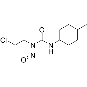 |
司莫司汀
CAS:13909-09-6 |
C10H18ClN3O2 |
|
Control of bull sperm cell volume during epididymal maturati...
2009-01-01 [Reprod. Fertil. Dev. 21(3) , 469-78, (2009)] |
|
Surveillance renal transplant biopsies and subclinical rejec...
2007-08-01 [Pediatr. Transplant. 11(5) , 536-9, (2007)] |
|
National Surgical Adjuvant Breast and Bowel Project trials i...
2001-02-01 [Semin. Oncol. 28(1 Suppl 1) , 9-13, (2001)] |
|
Quantification of meCCNU-induced dG-dC crosslinks in oligonu...
2011-07-30 [Rapid Commun. Mass Spectrom. 25(14) , 2027-34, (2011)] |
|
ANCA vasculitis: time for a change in treatment paradigm? No...
2011-06-01 [Rheumatology (Oxford.) 50(6) , 1019-24, (2011)] |