| 结构式 | 名称/CAS号 | 全部文献 |
|---|---|---|
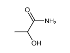 |
DL-乳酰胺
CAS:65144-02-7 |
| 结构式 | 名称/CAS号 | 全部文献 |
|---|---|---|
|
|
DL-乳酰胺
CAS:65144-02-7 |