| 结构式 | 名称/CAS号 | 全部文献 |
|---|---|---|
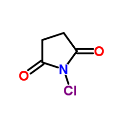 |
N-氯代丁二酰亚胺
CAS:128-09-6 |
|
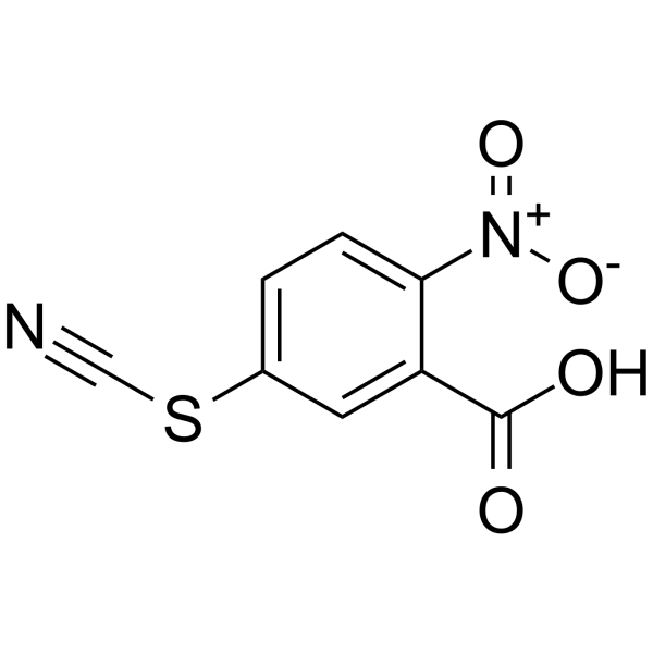 |
2-硝基-5-硫氰基苯甲酸
CAS:30211-77-9 |