| 结构式 | 名称/CAS号 | 全部文献 |
|---|---|---|
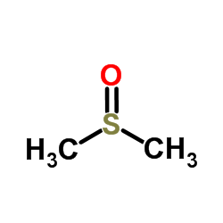 |
二甲基亚砜
CAS:67-68-5 |
|
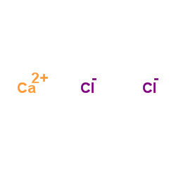 |
无水氯化钙
CAS:10043-52-4 |
|
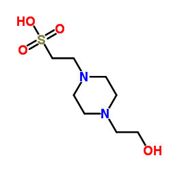 |
4-羟乙基哌嗪乙磺酸
CAS:7365-45-9 |
|
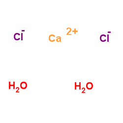 |
二水氯化钙
CAS:10035-04-8 |
|
 |
红海海绵素 B
CAS:76343-94-7 |