Capabilities of HPLC with APEX-Q nebulisation ICP-MS and ESI MS/MS to compare selenium uptake and speciation of non-malignant with different B cell lymphoma lines.
Heidi Goenaga-Infante, Shireen Kassam, Emma Stokes, Christopher Hopley, Simon P Joel
文献索引:Anal. Bioanal. Chem 399(5) , 1789-97, (2011)
全文:HTML全文
摘要
The formation of intracellular dimethylselenide (DMSe) as a product of exposure of non-malignant (PBMCs) and lymphoma (RL and DHL-4) cell lines to methylseleninic acid (MSA) at clinical levels is suggested here for the first time. This was achieved by analysis of cell lysates by HPLC coupled to ICP-MS via APEX-Q nebulisation, enabling limits of detection for target methyl-Se species which are up to 12-fold lower than those obtained with conventional nebulisation. Methyl-Se-glutathione (CH₃Se-SG), although detected in lysates of cells exposed to MSA, was found to be a reaction product of MSA with glutathione. This was confirmed by HPLC-ESI MS (MS) analysis of lysates of control cells (unexposed to Se) spiked with MSA. The MS/MS data obtained by collision-induced dissociation fragmentation of the ion m/z 402 (for [M+H](+) ⁸⁰Se) were consistent with the presence of CH₃Se-SG. Formation of DMSe was not detected by HPLC-ICP-MS in these spiked lysates, and it was found to require live cells in cell media containing MSA. Interestingly, the ratio of DMSe to CH₃Se-SG was significantly higher in lymphoma cells exposed to MSA in comparison to non-malignant cells. Moreover, maximum Se uptake levels in lymphoma cell lines seemed to be reached much earlier (after 10 min of MSA exposure) than in non-malignant cells. Finally, the GC-TOF-MS speciation data obtained for cell headspace suggested that the major Se species (dimethyldiselenide) appeared to be present in lymphoma cell headspace at significantly higher concentrations than in non-malignant cell headspace after only 10 min of exposure to MSA. Evidence for the presence of dimethylselenidesulfide in lymphoma cell headspace is also provided for the first time.
相关化合物
| 结构式 | 名称/CAS号 | 分子式 | 全部文献 |
|---|---|---|---|
 |
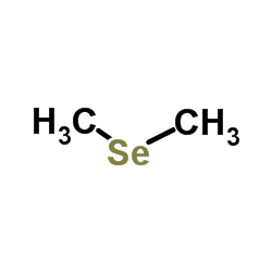
二甲基硒
CAS:593-79-3 |
C2H6Se |
|
Quantification of methylated selenium, sulfur, and arsenic i...
2014-01-01 [PLoS ONE 9(7) , e102906, (2014)] |
|
Gas chromatography with atomic emission detection for dimeth...
2005-11-18 [J. Chromatogr. A. 1095(1-2) , 138-44, (2005)] |
|
Biotransformation of L-selenomethionine and selenite in rat ...
2011-02-01 [Biol. Trace Elem. Res. 139(2) , 188-96, (2011)] |
|
First experimental observation of the CH3Se radical.
2005-10-14 [ChemPhysChem 6(10) , 2032-5, (2005)] |
|
Time-resolved study on the reactions of organic selenides wi...
2010-07-07 [Phys. Chem. Chem. Phys. 12(25) , 6750-8, (2010)] |