BNTA
Modify Date: 2025-08-26 11:30:09
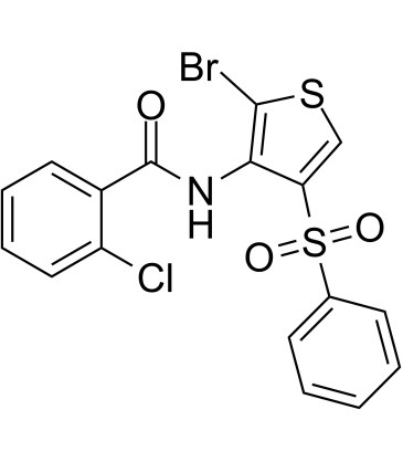
BNTA structure
|
Common Name | BNTA | ||
|---|---|---|---|---|
| CAS Number | 685119-25-9 | Molecular Weight | 456.76 | |
| Density | N/A | Boiling Point | N/A | |
| Molecular Formula | C17H11BrClNO3S2 | Melting Point | N/A | |
| MSDS | N/A | Flash Point | N/A | |
Use of BNTABNTA is a small molecule with extracellular matrix (ECM) modulatory properties, and it facilitates cartilage structural molecule synthesis on chondrocytes by activating superoxide dismutase 3 (SOD3). BNTA is a potential therapeutic agent in osteoarthritis[1]. |
| Name | BNTA |
|---|
| Description | BNTA is a small molecule with extracellular matrix (ECM) modulatory properties, and it facilitates cartilage structural molecule synthesis on chondrocytes by activating superoxide dismutase 3 (SOD3). BNTA is a potential therapeutic agent in osteoarthritis[1]. |
|---|---|
| Related Catalog | |
| In Vitro | BNTA (0.01-10 μM; 1-7 d) does not decrease cell viability of human osteoarthritis chondrocytes and rat primary chondrocytes[1]. BNTA (0.1 μM; 2 d) increases SOX9 protein markedly[1]. BNTA (0.1 μM; 2 d) remarkably increases the COL2A1 and SOX9 protein levels in IL1β-induced rat OA chondrocytes[1]. BNTA (10 μM; 5 d) increases proteoglycan staining in ATDC5 cells[1]. BNTA (0.01-10 μM; 6 h) upregulates the expression levels of ECM-related genes COL2A1, ACAN, proteoglycan 4 (PRG4), and SRY-box 9 (SOX9) in human OA chondrocytes[1]. BNTA (0.01-10 μM; 6 h) increases Col2a1, Acan, Prg4, and Sox9 mRNA levels, with maximum effects around 0.1 μM in IL1β-induced rat OA chondrocytes[1]. BNTA (0.01-1 μM; 2 or 3 w) enhances anabolism and inhibited inflammatory response in osteoarthritis cartilage explants[1]. Cell Viability Assay[1] Cell Line: Human OA chondrocytes Concentration: 0.01, 0.1, 1, 10 μM Incubation Time: 1, 3, 5, 7 d Result: No toxicity was observed. Western Blot Analysis[1] Cell Line: Human OA chondrocytes Concentration: 0.1 μM Incubation Time: 2 d Result: Elevated SOX9 protein compared with vehicle. |
| In Vivo | BNTA (0.015-1.5 mg/kg; intra-articular injection; twice a week for 4 and 8 weeks) could attenuate OA progression developed after anterior cruciate ligament transection (ACLT) in rats[1]. Animal Model: Male SD rats weighing 80 g are induced by ACLT[1] Dosage: 0.015, 0.15, 1.5 mg/kg Administration: Intra-articular injection; twice a week for 4 and 8 weeks Result: Attenuated post-traumatic osteoarthritis development after intra-articular injection for 4 and 8 weeks and was well tolerated. |
| References |
| Molecular Formula | C17H11BrClNO3S2 |
|---|---|
| Molecular Weight | 456.76 |