| Description |
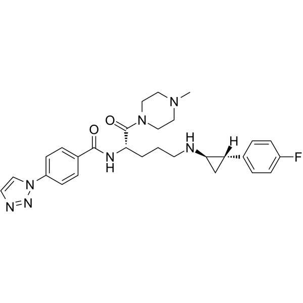
Bomedemstat (IMG-7289) is an oral and irreversible inhibitor of the epigenetically active lysine-specific demethylase 1 (LSD1) in mouse models of myeloproliferative neoplasms (MPNs). Bomedemstat can be used for the research of acute myelogenous leukemia (AML) and myelofibrosis (MF). Antineoplastic activity[1].
|
| Related Catalog |
|
| In Vitro |
Bomedemstat (IMG-7289) selectively inhibits proliferation and induced apoptosis of JAK2V617F cells by concomitantly increasing expression and methylation of p53, and, independently, the pro-apoptotic factor PUMA and by decreasing the levels of its antiapoptotic antagonist BCL-XL[1]. Bomedemstat (25 nM, 50 nM) and Ruxolitinib (175 nM) synergize in inhibiting JAK2V617F-driven proliferation[1]. Bomedemstat (50 and 100 nM) exerts a pro-apoptotic effect on 3 key regulators of programmed cell death, TP53, BCL-XL, and PUMA[1]. Cell Viability Assay[1] Cell Line: The human cell lines SET-2 (ATCC 608) and HEK293 Concentration: 25 nM, 50 nM Incubation Time: 96 hours Result: 25 nM alone significantly reduced colony formation. Western Blot Analysis[1] Cell Line: SET-2 cells Concentration: 50 and 100 nM Incubation Time: Result: Decreased levels of the antiapoptotic protein BCL-XL and increased levels of the pro-apoptotic protein PUMA.
|
| In Vivo |
Once-daily treatment with Bomedemstat (IMG-7289; 45 mg/kg) normalizes or improves blood cell counts, reduces spleen volumes, restores normal splenic architecture, and reduces bone marrow fibrosis[1]. Animal Model: Mx1cre-Jak2V617F mice[1] Dosage: 45 mg/kg Administration: Administered daily by oral gavage for either 14, 42, or 56 consecutive days Result: In this Mx-Jak2V617F model of myeloproliferative neoplasm (MPN), mice developed severe splenomegaly (up to 10-fold increase in spleen weight). Splenic architecture was completely destroyed, eliminating demarcation of the white and red pulp. Treatment significantly reduced splenomegaly with a few treated mice normalizing their spleen weight. Remarkably, the 56-day course led to partial restoration of lymph follicles and spleen architecture by histological examination.
|
| References |
[1]. Jonas S Jutzi,et al. LSD1 Inhibition Prolongs Survival in Mouse Models of MPN by Selectively Targeting the Disease Clone. Hemasphere.2018 Jun 8;2(3):e54.
|
